Fig. 2
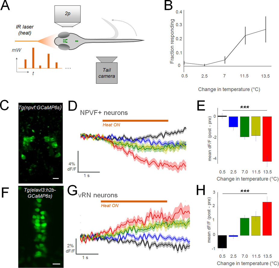
Noxious stimuli inhibit NPVF neurons and activate the vRN.
(A) Schematic of experimental configuration (mW: milliwatt, output of laser; t: time). (B) Results of behavior in wildtype larvae (n = 12; mean ± SEM). (C) 2p imaging field of view in Tg(npvf:GCaMP6s) larva: average of time-series. (D,G) Summed responses of all recorded neurons in each group. Colors indicate level of heat, as denoted in summary graphs (E,H) to right (±SEM). In a single larva, multiple cells were analyzed (10 trials in each heat level, 50 trials total per larva). NPVF: n = 113 neurons in 13 larvae. vRN neurons: 173 cells in 6 larvae. (E,H) Summary data for each cell type (±SEM). 1-way anova test. ***p < 0.001. (F) 2p imaging field of view in Tg(elavl3:h2b-GCaMP6s) larva: average of time-series. Scale bar: 10 μm.

