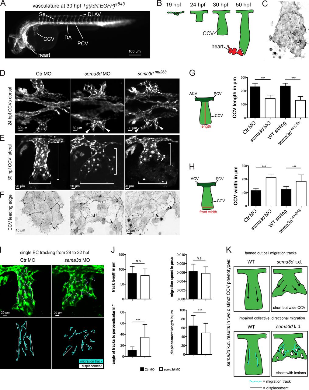
Fig. 1 Sema3d regulates collective EC migration by controlling CCV width and EC sheet organization. (A) Vasculature of a 30-hpf-old zebrafish embryo visualized by GFP expression of Tg(kdrl:EGFP)s843. (B) Illustration of CCV development. (C) CCV ECs migrate as a collective cell sheet; F-Actin visualized by GFP expression of Tg(fli1a:lifeactEGFP)mu240 at 32 hpf. (D–F) CCV development is impaired in sema3d morphants and mutants. (D) Widened CCV outgrowth upon Sema3d loss at 24 hpf (white arrowheads); confocal projections of Tg(kdrl:EGFP)s843 embryos. (E) Shorter but wider CCVs of sema3d morphants and mutants at 30 hpf (white brackets). (F) The CCV cell sheet exhibits lesions (black arrowheads) in the leading edges of 30-hpf Sema3d-deficient Tg(fli1a:lifeactEGFP)mu240 embryos. Confocal projections were color-inverted. (G) Quantification of CCV length shows a shortening in sema3d morphants and mutants at 30 hpf (each n = 25). (H) Quantification of CCV front width shows an increase in sema3d morphants and mutants at 30 hpf (each n = 18). (I and J) Tracking of single CCV EC migration from 28 to 32 hpf. (I) Confocal projections at 32 hpf with migration tracks (turquoise lines) and displacement distance (white arrows). (J) Quantification of migration parameters (each condition 84 cells and 14 cells per embryo/movie). Track length and migration speed are not impaired in sema3d morphants. In contrast, track displacement length is reduced and the angles of tracks to the perpendicular are increased in sema3d morphants compared with Ctr morphants, leading to a fanned-out CCV. (K) Model of sema3d knockdown (k.d.) representing the two distinct phenotypes: first, fanned-out cell migration tracks leading to a shorter but wider CCV; second, impaired collective and directional migration and a disrupted EC sheet. DA, dorsal aorta; ACV, anterior cardinal vein; PCV, posterior cardinal vein. ***, P < 0.001; n.s., not significant. Error bars indicate SD.
Image
Figure Caption
Figure Data
Acknowledgments
This image is the copyrighted work of the attributed author or publisher, and
ZFIN has permission only to display this image to its users.
Additional permissions should be obtained from the applicable author or publisher of the image.
Full text @ J. Cell Biol.

