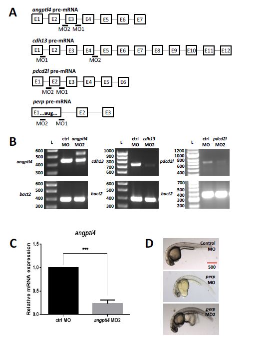
Fig. S10
Design and validation of second non-overlapping MOs to target angptl4, cdh13, pdcd2l and perp. (A) Diagramatic representation of angptl4, cdh13, pdcd2l and perp pre-mRNAs with exons (numbered boxes) and introns (solid lines). Exon and intron sizes are arbitrary. The regions in pre-mRNA where MOs bind are indicated with black bars labelled MO1 or MO2. (B) Zebrafish embryos were injected with second non-overlapping MO (MO2) for each gene of interest. The ability of angptl4 MO2, cdh13 MO2 and pdcd2l MO2 to modify splicing of the targeted transcript was assessed by analysis of RT-PCR products after agarose gel electrophoresis. Splicing modifications were observed as a band shift or as a reduction of the wildtype band compared to the control sample (ctrl MO). RT-PCR analysis of beta-actin (bact2) was used as an internal control. The DNA fragment sizes are indicated in base pairs next to the DNA ladder (L). (C) Embryos with residual expression of the wildtype transcript (angptl4 MO2 in (B)) were additionally tested by qPCR to validate MO efficiency. Mean values are shown with standard error of the mean from three independent experiments. ***p<0.001, using an unpaired two-tailed t-test. (D) Embryos injected with either perp MO or a non-overlapping MO (MO2) exhibited bursting of yolk cell during dechorionation using forceps, which was not observed in control embryos. Embryos are shown at 30 hpf, lateral view, anterior to the left, dorsal up. Scale bar: 500 μm.

