Fig. 1
- ID
- ZDB-IMAGE-170222-10
- Genes
- Antibodies
- Source
- Figures for Serbanovic-Canic et al., 2017
Fig. 1
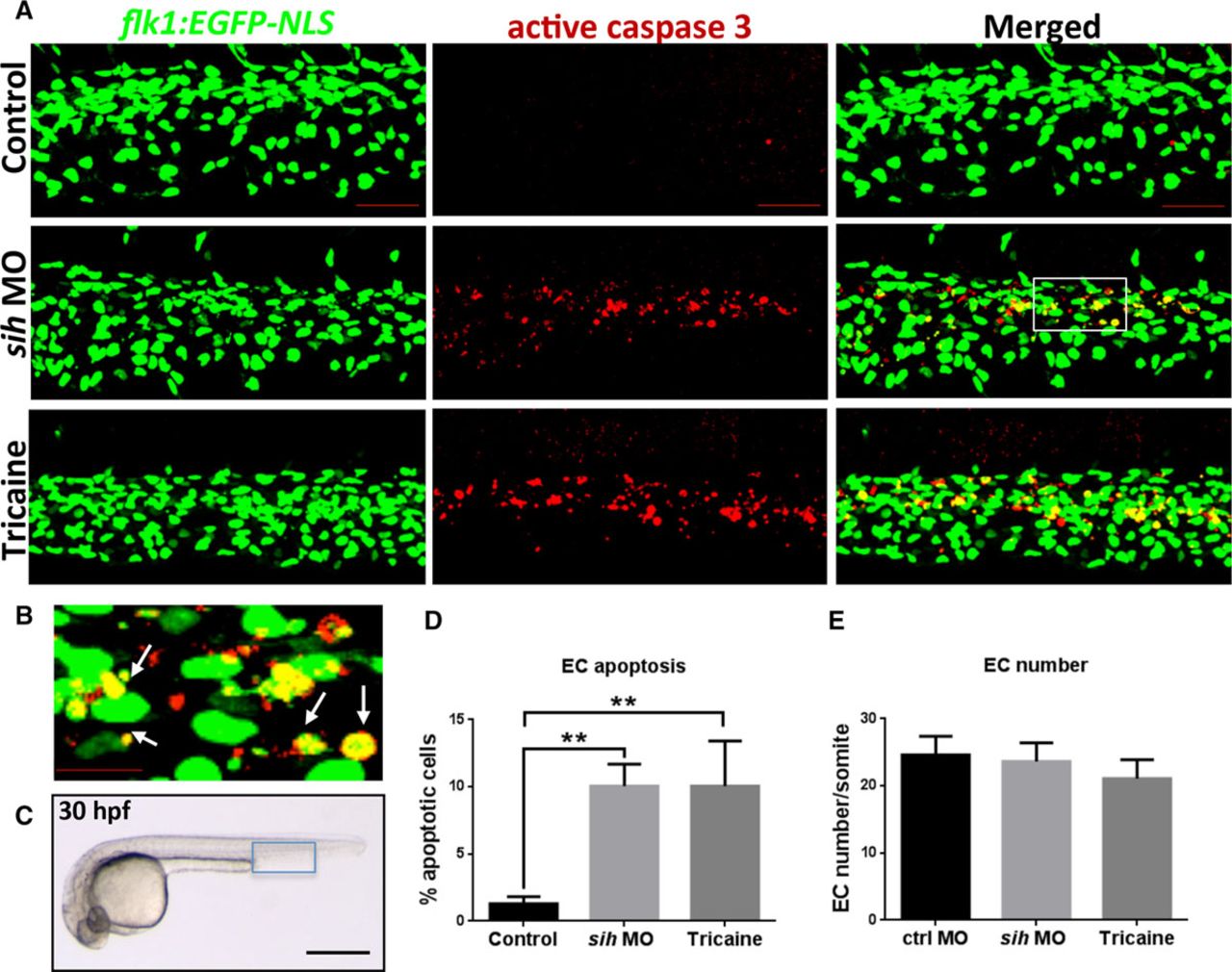
Flow cessation induces endothelial cell (EC) apoptosis in zebrafish embryos. A, Whole-mount active caspase-3 (red) staining of 30 hours post fertilization (hpf) flk1:EGFP-NLS zebrafish embryos (green EC nuclei) in the presence (control) or absence of flow (sih morpholino oligonucleotide [MO], tricaine). The region outlined with the white box is shown in higher magnification in B; white arrows indicate apoptotic ECs (yellow). C, Zebrafish embryo at 30 hpf. The region outlined with blue box represents the region that is studied in A. The percentage of EC apoptosis (D) and EC numbers (E) in sih MO-injected and tricaine-treated embryos compared with controls was quantified, and mean values are shown with SD; n≥15 from 3 independent experiments, **P<0.01 using 1-way ANOVA. A–C, Lateral view, anterior to the left, dorsal up. Scale bars, 50 μm (A), 15 μm (B), and 500 μm (C).

