
|
Fig. S2
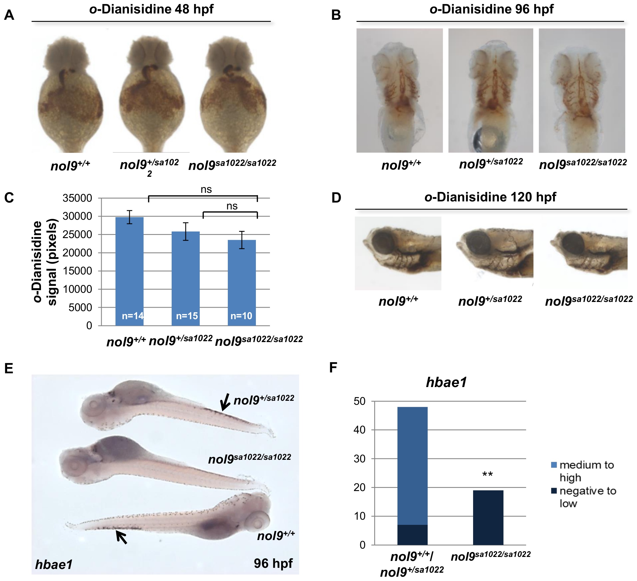
Definitive, but not primitive erythropoiesis is affected in nol9sa1022/sa1022 mutants.
(A-D) o-Dianisidine staining of circulating erythrocytes at 48 hpf, 96 hpf and 120 hpf. (A) Representative images of o-Dianisidine-stained embryos at 48 hpf. The level of staining was similar between nol9+/+ (n = 13), nol9+/sa1022 (n = 35) and nol9sa1022/sa1022 (n = 8) embryos. Ventral view with anterior up. (B) Representative images of o-Dianisidine-stained larvae at 96 hpf. The number of stained circulating primitive erythrocytes was comparable between nol9sa1022/sa1022 (n = 10), nol9+/+ (n = 15) and nol9+/sa1022 (n = 15) siblings. Ventral view with anterior up. (C) Quantification of the area stained in o-Dianisidine-stained larvae area at 96 hpf. Data are represented as average +/- SEM. Two-tailed Student’s t test, p>0.05. ns–not significant. (D) Representative images of o-Dianisidine-stained larvae at 120 hpf. All nol9sa1022/sa1022 (n = 20), nol9+/+ (n = 2) and nol9+/sa1022 (n = 8) larvae displayed a similar level of staining. Larvae oriented with anterior to the left and dorsal to the top. (E) Whole-mount in situ hybridization using hbae1 riboprobe at 96 hpf. Arrows indicate hbae1-positive definitive erythrocytes present in the CHT of nol9+/sa1022 and nol9+/+ but not in nol9sa1022/sa1022 larvae. (F) Quantification of hbae1 in situ hybridization data. Data are represented as the number of larvae belonging to each phenotypic group. Fisher’s exact test, **, p<0.01.