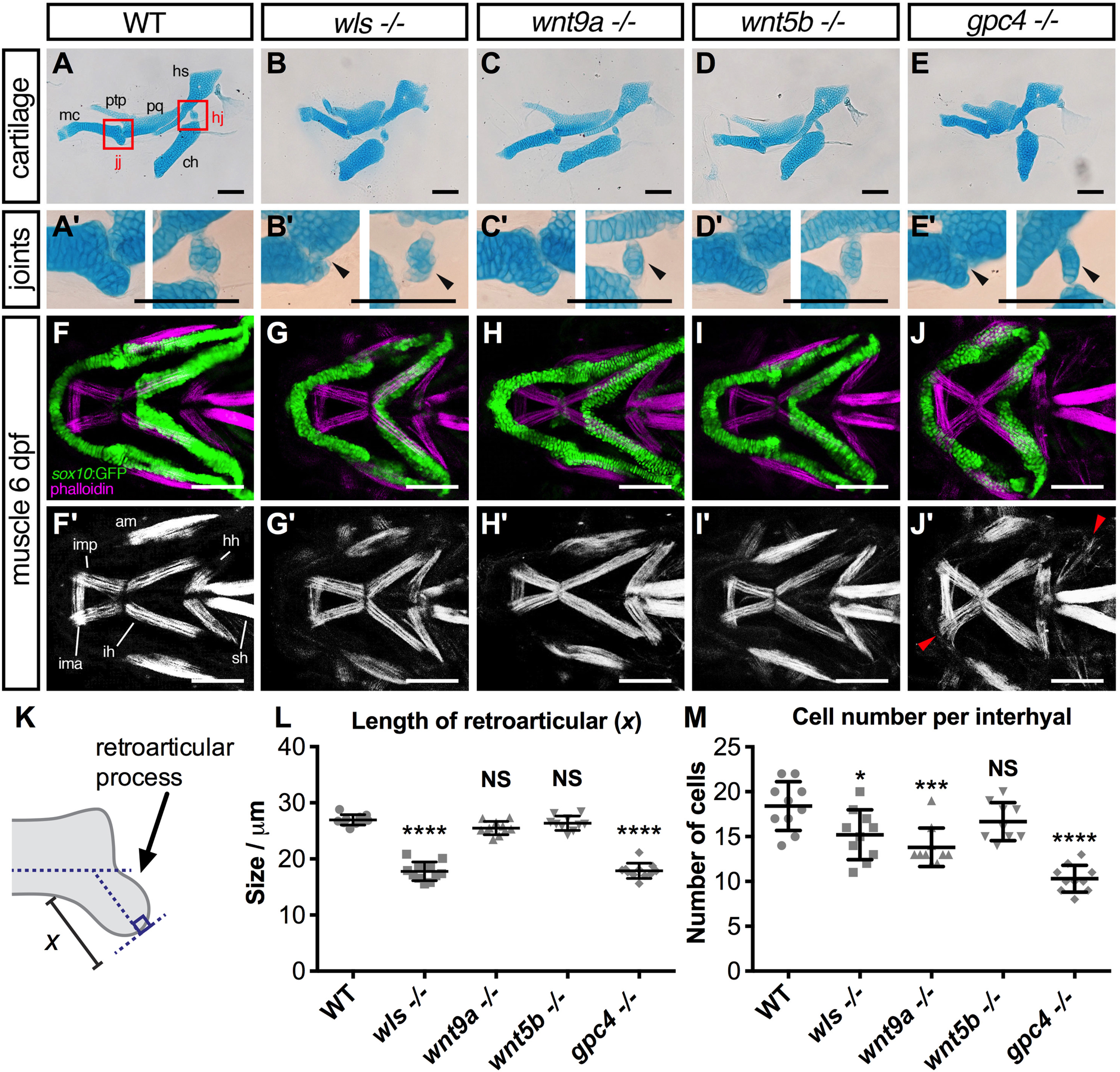
Fig. 5
Joint and muscle defects inwls,wnt9a,wnt5b, andgpc4mutants. (A-E) Alcian blue stained ventral cartilage lateral whole-mounts of 8 dpf embryos and 40X zoomed image in A′-E′ of the jaw joint (jj; left) and hinge joint (hj; right). Meckel's cartilage (mc), pterygoid process (ptp), palatoquadrate (pq), hyosymplectic (hs), and ceratohyal (ch). Jaw joint defects evident in wls and gpc4 mutant whilst wnt9a exhibited a slightly malformed retroarticular process. wls displayed a clump of cells within the hinge-joint compared to the neatly stacked layered chondrocytes in wild-type. In contrast, wnt9a, wnt5b and gpc4 were only a single cell layer in thickness. (F-J) Maximum intensity projections of 20X zoom confocal stacks showing muscle stain with phalloidin (purple) and anti-GFP (green) for cartilage elements. Muscle length defects apparent in all Wnt mutants with gpc4 displaying disorganized intermandibularis posterior (imp) invading the gaps in Meckel's cartilage. Adductor mandibulae (am), intermandibularis anterior (ima), intermandibularis posterior (imp), hyohyoideus (hh), interhyoideus (ih), sternohyoides (sh) F′-J’’ Single channel phalloidin flourescence. (K) Graphical representation of retroaritcular measurement. (L) Measured length of retroarticular process and (M) cell count per interhyal in WT and Wnt mutants. (*p<0.01***p<0.001, ****p<0.0001; Kruskal-Wallis test with Dunn's multiple comparison test. Significance level at p<0.05). Scale =50 µm..
Reprinted from Developmental Biology, 421(2), Ling, I.T., Rochard, L., Liao, E.C., Distinct requirements of wls, wnt9a, wnt5b and gpc4 in regulating chondrocyte maturation and timing of endochondral ossification, 219-232, Copyright (2017) with permission from Elsevier. Full text @ Dev. Biol.

