Fig. 4
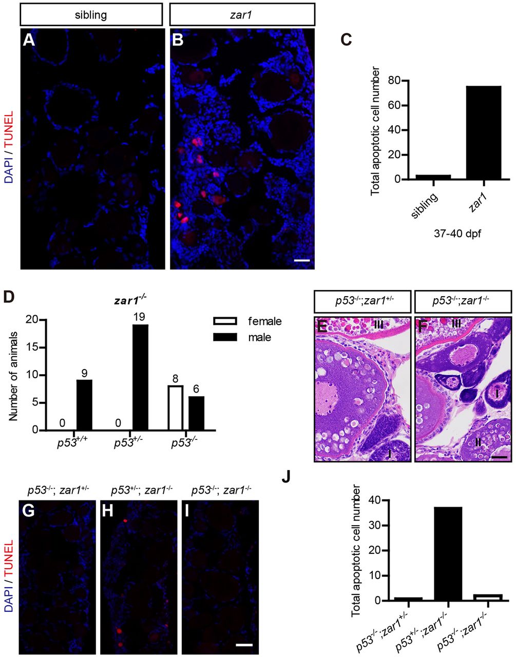
Zar1 deficiency causing all-male phenotype is due to p53-mediated apoptosis. (A-C) TUNEL staining of ovary sections of zar1 homozygotes and control siblings at 37-40 dpf. Obvious apoptotic cells are observed in immature ovaries in zar1 homozygotes (A), but not in sibling controls (B). Quantification of apoptotic cells is shown in C; 18 sections from 6 juveniles were counted for each genotype. (D) Gender analysis of zar1−/− homozygotes on different p53 genotype backgrounds (p53+/+, p53+/− and p53−/−). Females are only observed in p53−/−;zar1−/− double homozygous mutants. (E,F) H&E staining of sections of p53−/−;zar1+/− and p53−/−;zar1−/− adult ovaries. p53−/−;zar1−/− ovaries are morphologically normal. (G-J) TUNEL staining of ovary sections of p53−/−;zar1−/− ovaries at 37-40 dpf with p53−/−;zar1+/− and p53+/−;zar1−/− ovaries as controls. (J) Quantification of apoptotic cells. Nine sections from three juveniles were counted for each genotype. Scale bar: 20 μm.

