Fig. 4
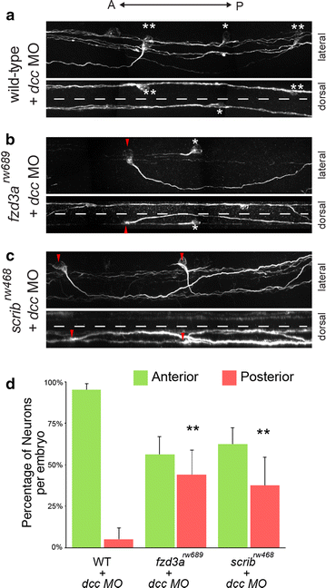
Anterior guidance of CoPA axons in the absence of midline crossing is dependent on PCP proteins. a CoPA axons fail to cross the midline in dcc knockdown embryos yet still undergo anterior growth. In the lateral view, asterisk indicates affected CoPA that pathfinds anteriorly with no ventral growth. Double asterisk indicates affected CoPA that displays weak ventral extension followed by projection dorsally and anteriorly without crossing the midline. Dorsal view of the same spinal cord demonstrates that affected CoPAs are prevented from crossing the midline (dotted line), b, c CoPA axons in fzd3a rw689 and scrib rw468 embryos injected with dcc morpholinos also fail to cross the midline. Red arrowhead indicates uncrossed CoPA axons that grow posteriorly inappropriately in dcc-deficient fzd3a rw689 and scrib rw468 embryos. Asterisk indicates uncrossed CoPA axon that projects anteriorly. Dorsal views of the same spinal cords demonstrate that posteriorly projecting CoPAs fail to cross the midline (dotted line), d quantification of the anterior or posterior trajectory of uncrossed CoPA neurons in dcc-deficient WT, fzd3a rw689 , and scrib rw468 embryos. Error indicates SD. **p < 0.01 versus WT + dcc MO

