Fig. S6
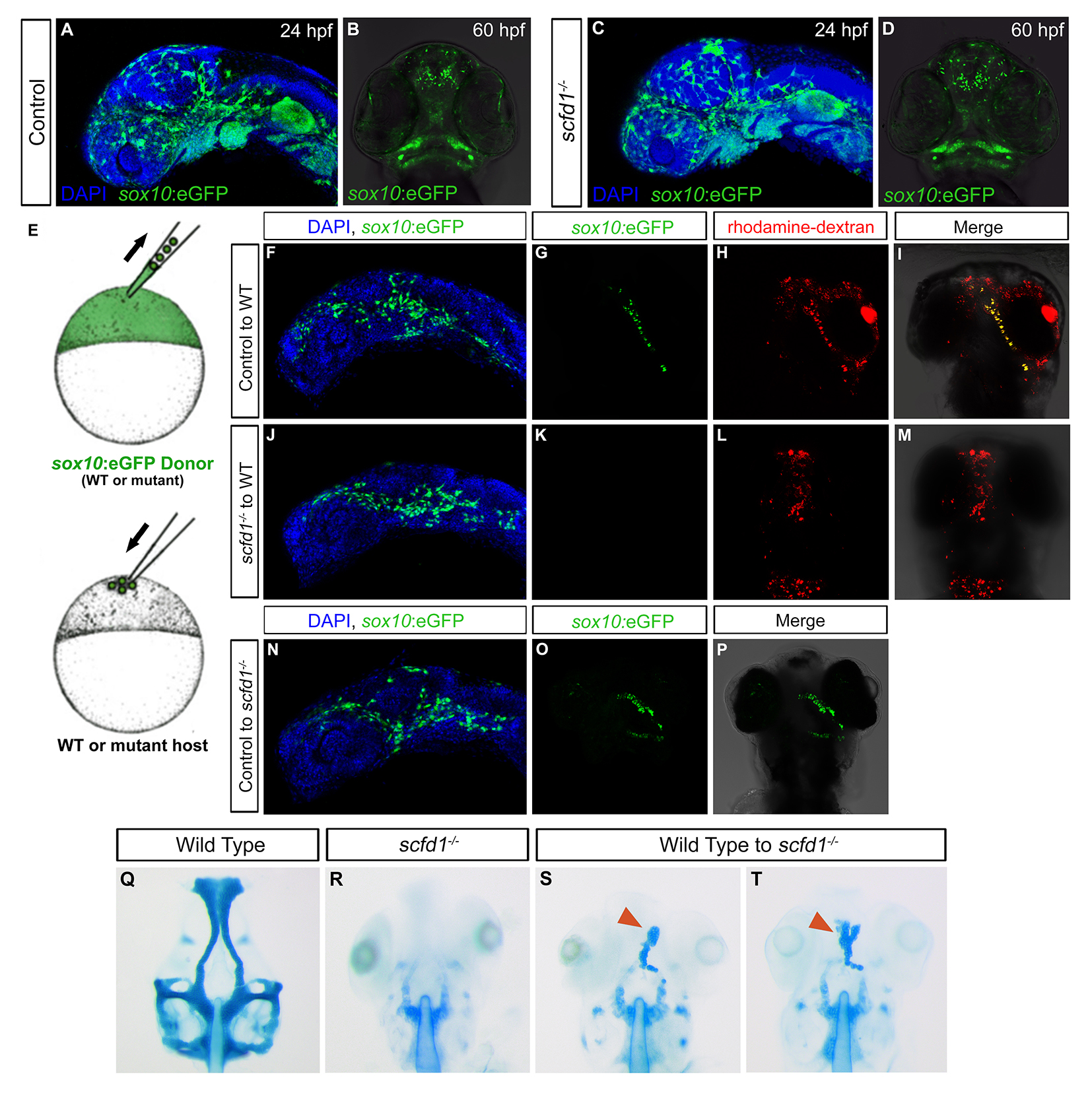
Cell autonomous requirement for scfd1 in neural crest cells for cartilage differentiation. (A-D) Sox10: eGFP transgene expression in control and scfd1 mutant embryo. A and C, lateral views with anterior to left. B and D, ventral views with anterior to the top. (E) Schematic diagram of the transplantation approach. At 4 hpf, cells from a donor (wild type or mutant) Sox10:eGFP transgenic embryo are transplanted to the animal pole of a wild type or scfd1 mutant host embryo. (F, J and N) At 24 hpf, donorderived neural crest cell migration to the oral ectoderm is evident regardless of donor genotype. Lateral views with anterior to the top. (G-I) At 72hpf, in control transplantation experiments, donor cells were observed contributing to cartilage. (J-M) Conversely, in none of the transplants where scfd1 mutant donor cells were used was contribution to cartilage noted. (N-P) We also observed that wild type donor GFP-positive neural crest cells in scfd1 mutant host embryos formed cartilage. (Q-T) At 72 hpf, cartilage staining reveals wild type donor-derived cells partially rescuing anterior neurocranium defects in scfd1 mutant embryos. Red arrowheads indicate cartilage derived from wild type donor cells.
Reprinted from Developmental Biology, 421(1), Hou, N., Yang, Y., Scott, I.C., Lou, X., The Sec domain protein Scfd1 facilitates trafficking of ECM components during chondrogenesis, 8-15, Copyright (2017) with permission from Elsevier. Full text @ Dev. Biol.

