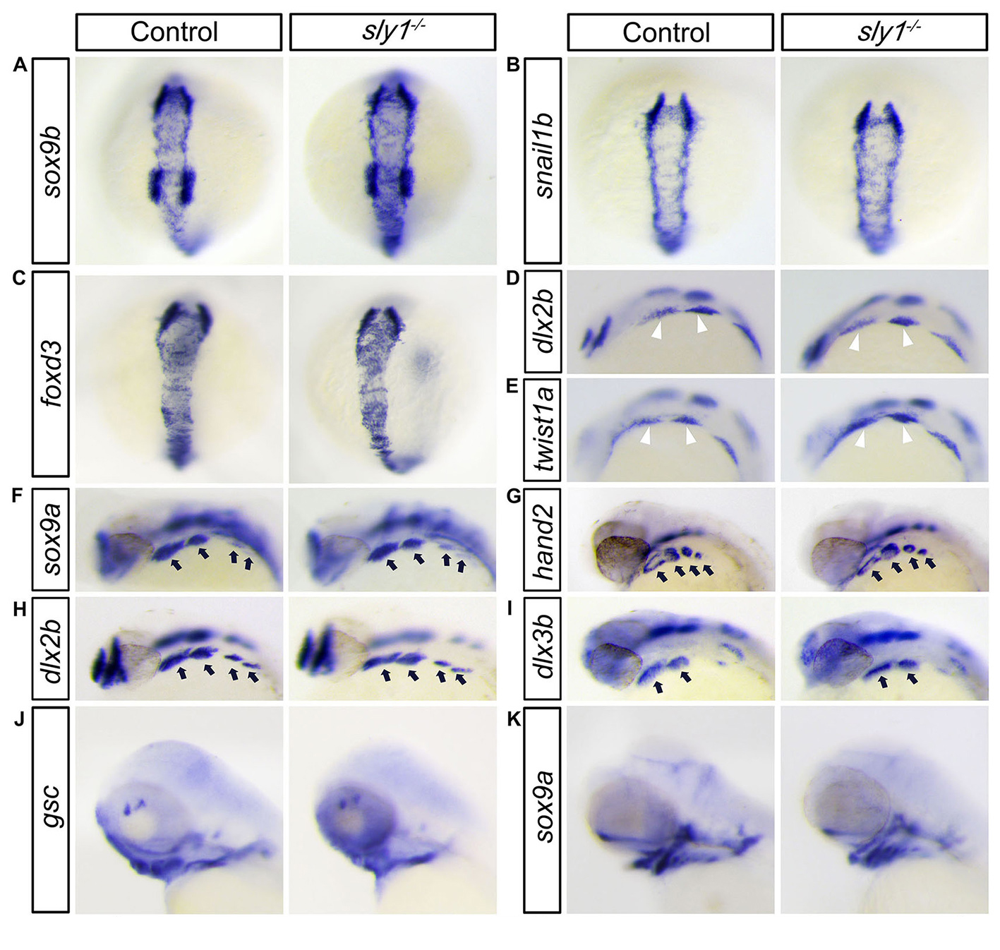
Fig. S5
In scfd1 mutant embryos, development of chondrogenic neural crest cell appears normal until initiation of chondrogenesis. (A-C) Sox9b, foxd3, and snail1b are expressed in pre-migratory neural crest at the dorsal side of neural tube at 14 hpf. No overt differences in expression were evident via RNA in situ hybridization between control and mutant embryos, indicating that neural crest cells were properly specified in scfd1 mutant embryos. (D-E) RNA in situ hybridization showed that markers of migratory facial neural crest (white arrowhead), dlx2a and twist1a, were expressed normally in mutants at 18-somite stage (18 hpf). D and E, lateral view with anterior to the top. (F-I) RNA in situ hybridization showed that sox9a, hand2, dlx2b and dlx3b, a group of gene controlling pharyngeal arch (black arrows) patterning and outgrowth, were expressed normally in scfd1 mutants at 32 hpf. D to K, dorsal views with anterior to the top. (J-K) The expression patterns of goosecoid and sox9a, regulators of mesenchymal condensation and chondrocyte differentiation, were unaffected in scfd1 mutants until 72 hpf, indicating that chondrogenesis is initiated normally in scfd1 mutants. L and K, lateral views with anterior to the left.
Reprinted from Developmental Biology, 421(1), Hou, N., Yang, Y., Scott, I.C., Lou, X., The Sec domain protein Scfd1 facilitates trafficking of ECM components during chondrogenesis, 8-15, Copyright (2017) with permission from Elsevier. Full text @ Dev. Biol.

