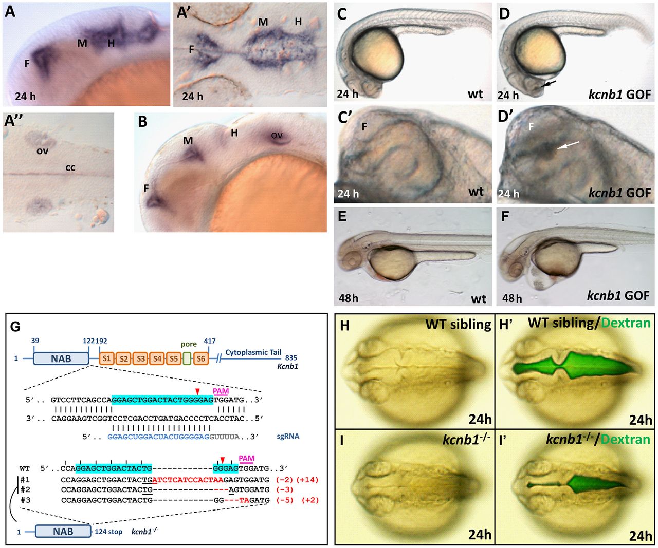
Fig. 7
kcnb1 is antagonistic to kcng4b when expressed in the brain. (A-B) Antisense Dig-RNA WISH detected kcnb1 transcripts in the ventricular system at 24 and 30 hpf. (A,B) Lateral views with anterior to the left. (A′,A″) Flat mounts of WISH-stained embryo with anterior to the left. (C,D) kcnb1 GOF 24 hpf embryos contain a clump of cells (arrow) in the third ventricle (lateral view, anterior to the left). (C′,D′) Zoomed image of the forebrain with a clump of cells (arrow). (E,F) At 48 hpf, embryos develop hydrocephalus and cardiac edema. (G) Schematic illustration of kcnb1 mutant generated using CRISPR-Cas system. Position of the target site, target sequence (blue), single guide RNA (sgRNA) sequence, PAM location (pink), isolated indel mutants (indels in red) and their respective sequences and mutant with truncated kcnb1 polypeptide are shown. In mutants #1, #2, a premature stop codon (TGA) was found. Mutant #3 is a mis-sense mutant. Mutant #1 was used for all subsequent analysis. (H,I) kcnb1 LOF results in under-inflated ventricles. (H′,I′) Ventricles filled with 70 kDa FITC-Dextran. Dorsal views with anterior to the left. cc, central canal; F, forebrain; H, hindbrain; M, midbrain; ov, otic vesicle.

