Fig. 1
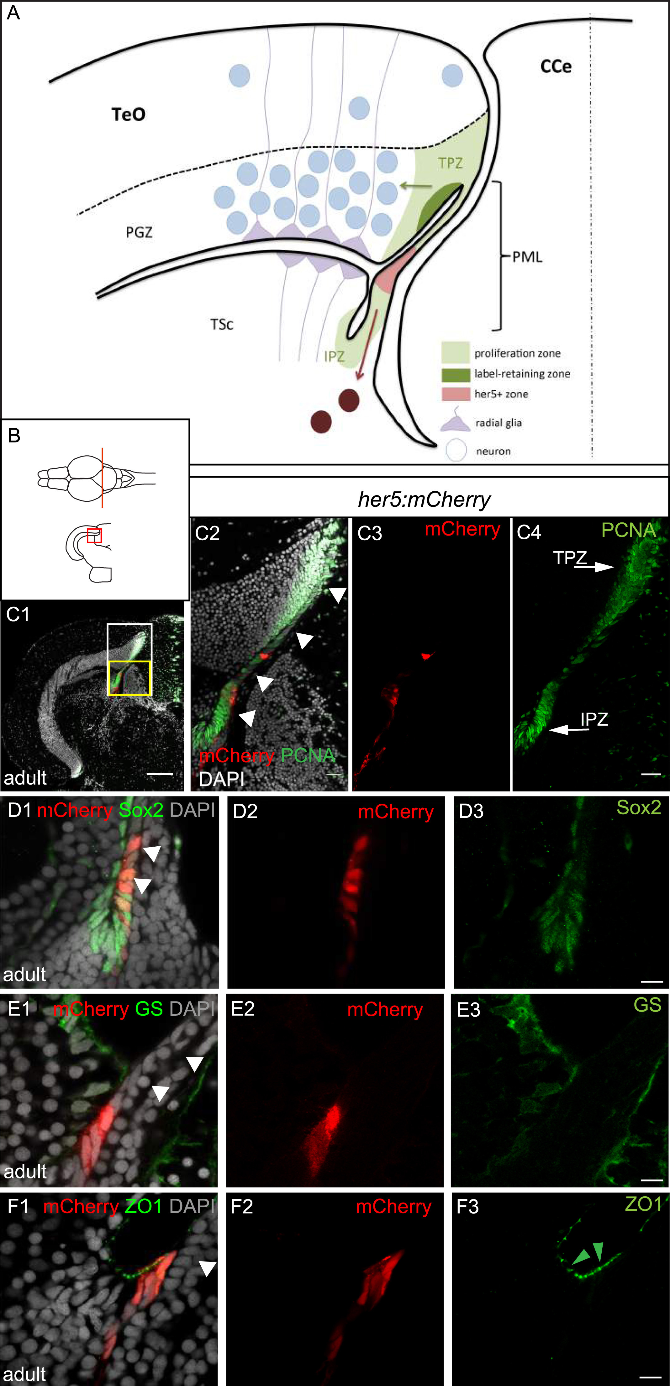
Adult her5-positive cells of the PML express the characteristics of neuroepithelial progenitors. A, B: Schematic cross-section of one tectal hemisphere from an adult zebrafish (level indicated in B, dorsal up, midline indicated by the vertical broken line) showing the different relevant cell populations (color-coded, see keys on figure). Arrows indicate the known lineages, ie: green arrow: generation of PGZ neurons and radial glia from TPZ proliferating progenitors ( Alunni et al., 2010 and Ito et al., 2010); dark red arrow: generation of tegmental neurons by her5-positive cells of the PML ( Chapouton et al., 2006). C: Cross section of an adult optic tectum at the level indicated in B, immunostained for mCherry and PCNA (color-coded) and counterstained with DAPI, position of the PML (arrowheads) and the Her5-mCherry-positive domain compared to the TPZ and IPZ. C1: low magnification showing one hemisphere, C2-C4: high magnifications of the white boxed area in C1 (C2: red and green channels with DAPI, C3: red channel only, C4: green channel only). Arrowheads to the PML on panel C2. D-F: compared expression of Her5-mCherry with markers for progenitors (Sox2), glial cells (GS) and tight junctions in neuroepithelial cells (ZO1, green arrowheads) on high magnifications of the PML area on cross sections of an adult optic tectum (C1-F3: High magnification of the yellow boxed area in C1, panels 1: red and green channels with DAPI, panels 2: red mCherry channel only, panels 3: green channel only). White arrowheads to the PML on panels 1. Scale bars: C 50 µm, D-F 10 µm. Abbreviations: CCe: crista cerebellaris, IPZ: isthmic proliferation zone, PGZ: periventricular grey zone, PML: peripheral midbrain layer/posterior midbrain lamina, TeO: tectum opticum, TPZ: tectal proliferation zone, TSc: torus semi-circularis.
Reprinted from Developmental Biology, 420(1), Galant, S., Furlan, G., Coolen, M., Dirian, L., Foucher, I., Bally-Cuif, L., Embryonic origin and lineage hierarchies of the neural progenitor subtypes building the zebrafish adult midbrain, 120-135, Copyright (2016) with permission from Elsevier. Full text @ Dev. Biol.

