
|
Fig. S7
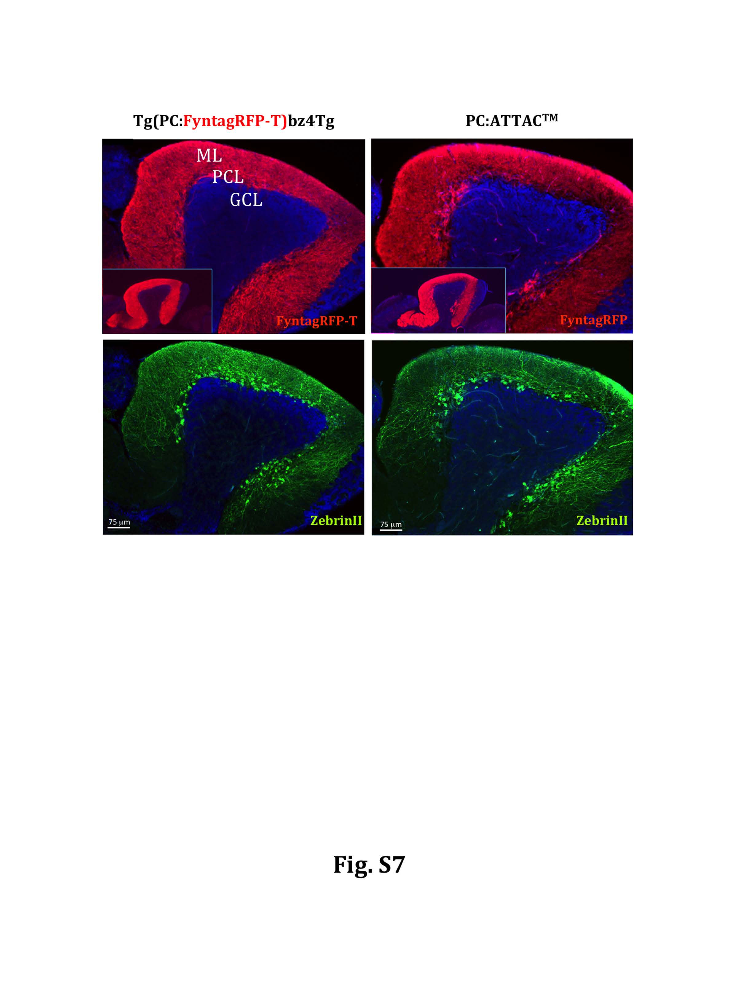
Integrity of Purkinje cell layer in PC-ATTACTM adults. Immunohistchemical detection of Purkinje cell specific FyntagRFP and ZebrinII expression on sagittal sections through the cerebellum of adult fish older than 15 months from the control Tg(PC:FyntagRFP-T)bz4Tg (left) and the PC-ATTACTM (right) strains. Both lines showed intensive FyntagRFP-T/FyntagRFP expression respectively without any signs of dendritic atrophy (upper row, red fluorescence, insets display overview over entire cerebellum), and ZebrinII expression (lower row, green fluorescence) revealed that no apparent Purkinje cell loss was observed in the PC-ATTACTM cerebellum compared to controls. DAPI was used as counter stain (blue fluorescent signals). Abbr.: GCL: granule cell layer, ML: molecular layer, PCL: Purkinje cell layer. Scale bar: 75μm.