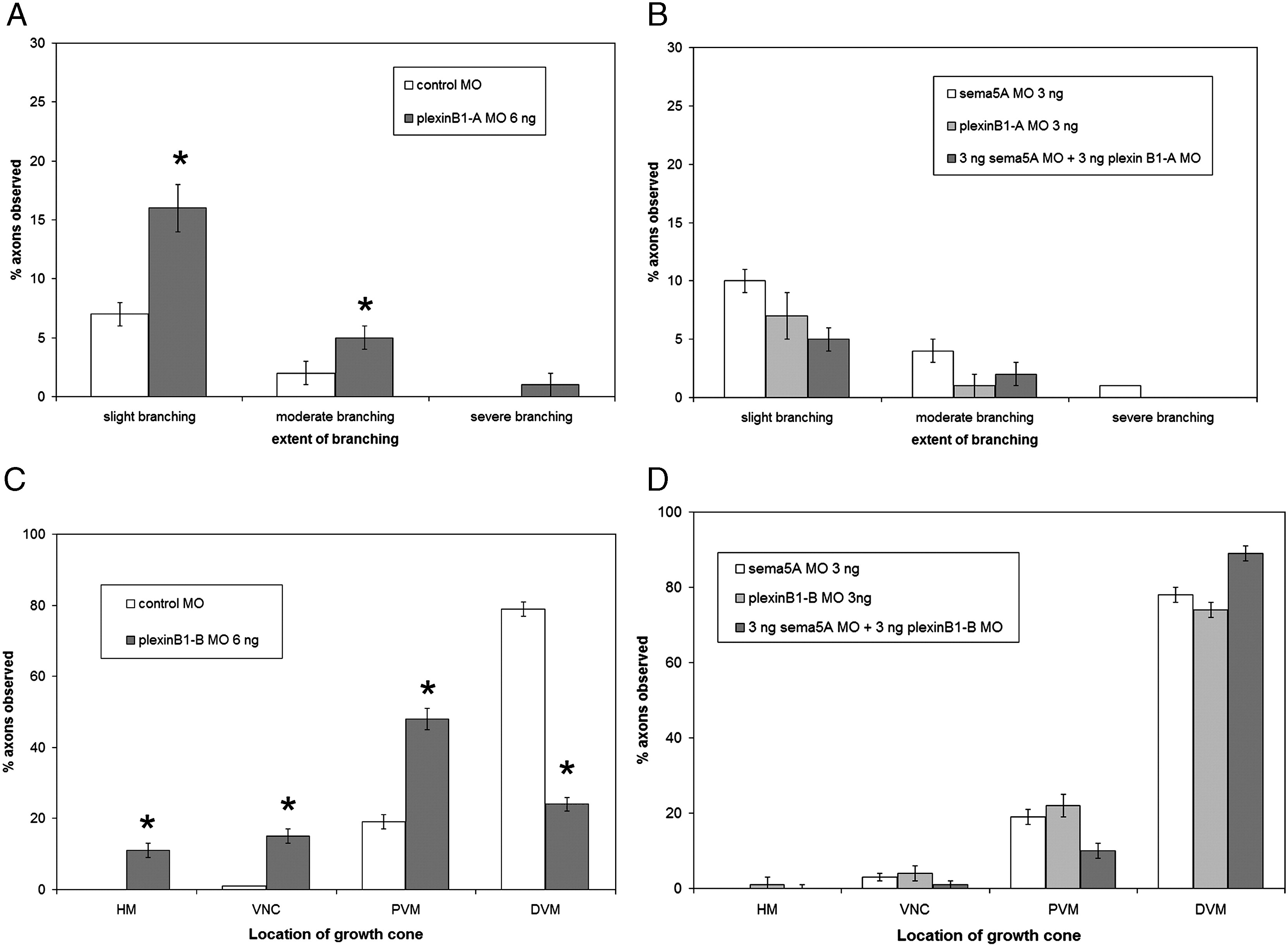
Fig. 4
plexin B1-A and plexin B1-B morpholino injection phenotypes and co-injections with sema5A. (A) Injection of 6 ng plexin B1-A MO shows an increase in branching CaP axons compared to control MO injected embryos. (B) Co-injection of 3 ng plexin B1-A MO and sema5A MO has little or no difference with injection of each MO by itself at 3 ng. (C) Injection 6 ng plexin B1-B MO results in a delay in CaP axon extension into the ventral myotome compared control MO injected embryos. (D) Co-injection of 3 ng of plexin B1-B MO and sema5A MO displayed no delay of CaP axon extension phenotype. Data quantified from 3 ng sema5A MO injected embryos (n = 2020 axons, 101 embryos), 6 ng plexin B1-A MO injected embryos (n = 1040 axons, 52 embryos), 3 ng plexin B1-A MO injected embryos (n = 940 axons, 47 embryos), 3 ng sema5A MO + 3 ng plexin B1-A MO injected embryos (n = 2140 axons, 107 embryos), 6 ng plxnB1B MO injected embryos (n = 1960 axons, 98 embryos), 3 ng plexin B1-B MO injected embryos (n = 1080 axons, 54 embryos), 3 ng sema5A MO + 3 ng plexin B1-B MO injected embryos (n = 1180 axons, 59 embryos). Error bars represent confidence interval for proportions at 95% confidence. Asterisks indicate significant difference between control MO injected and plexin B1-A or plexin B1-B MO injected with p < 0.001 using one-way ANOVA.
Reprinted from Developmental Biology, 326(1), Hilario, J., Rodino-Klapac, L.R., Wang, C., and Beattie, C.E., Semaphorin 5A is a bifunctional axon guidance cue for axial motoneurons in vivo, 190-200, Copyright (2009) with permission from Elsevier. Full text @ Dev. Biol.

