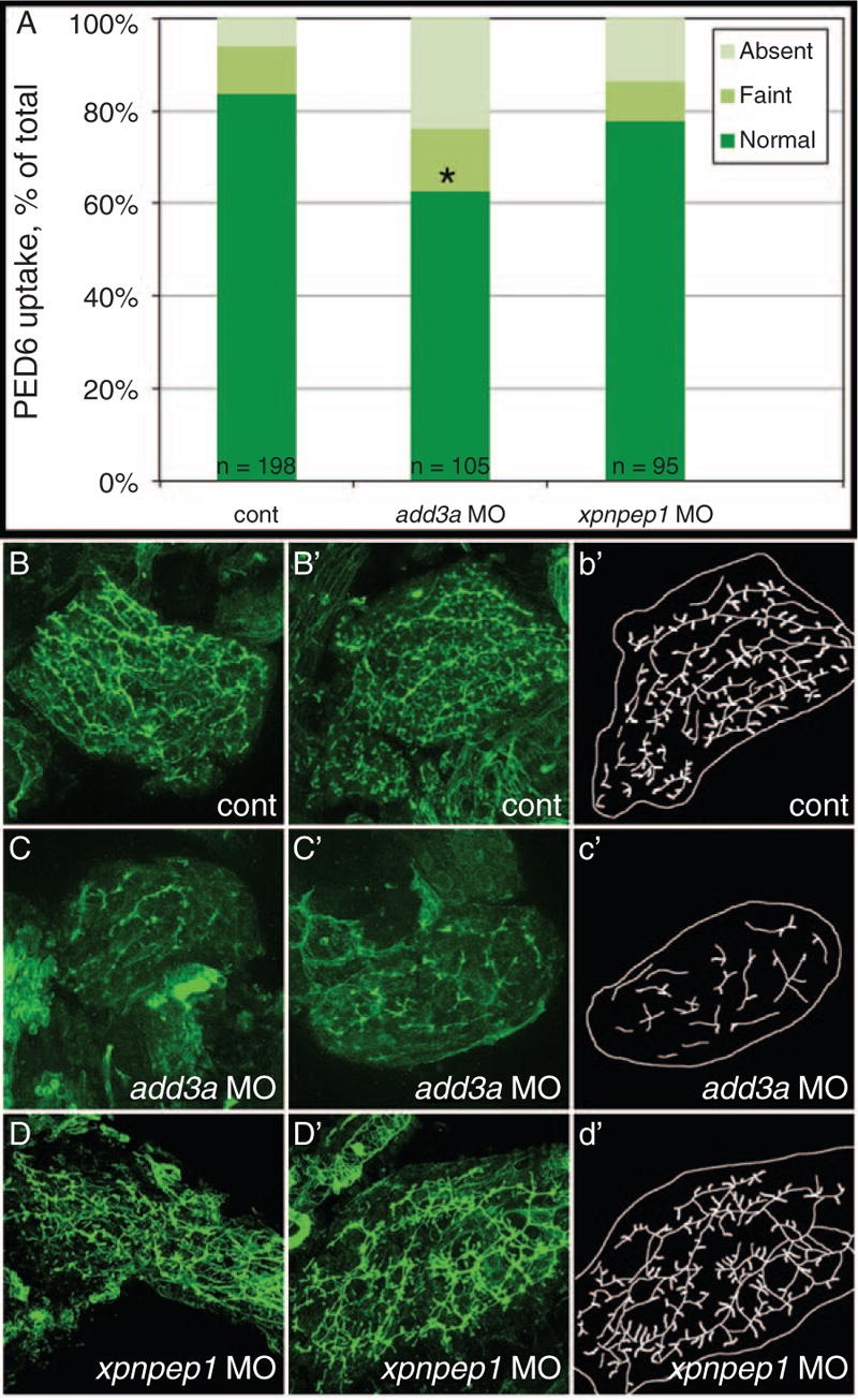
Fig. 2
Decreased biliary function and developmental biliary abnormalities in add3a morpholino-injected 5 days post fertilization (dpf) larvae. Wild-type fertilized oocytes were injected with MO directed against add3a or xpnpep1, or control. At 5 dpf, larvae were incubated in the fluorescent lipid PED6 and assayed for gallbladder uptake, and then fixed and stained for cytokeratin to document biliary anatomy. A, Percentages of control (cont), add3a MO-injected and xpnpep1 MO-injected larvae with normal, faint, or absent gallbladder visibility, reflecting PED6 uptake and thus biliary function. There is a significant decrease in biliary function in the add3a MO-injected larvae (*P ≤ 0.0001 by chi-square analysis), whereas the difference between control and xpnpep1 MO-injected larvae is not significant. B, Confocal projections of cytokeratin immunostainings of livers from 5 dpf larvae injected with control (cont, B), add3a MO (C), or xpnpep1 MO (D). There are 2 examples of each condition (B, B'; C, C'; D, D'), and a schematic outline of each second example (b', c', d'). Note that the pattern of the intrahepatic ducts from the cont and xpnpep1 MO-injected larvae appears similar, whereas the add3a MO-injected larvae have shorter and sparser ducts. Original magnification ×400. MO = morpholino antisense oligonucleotide.

