Fig. S5
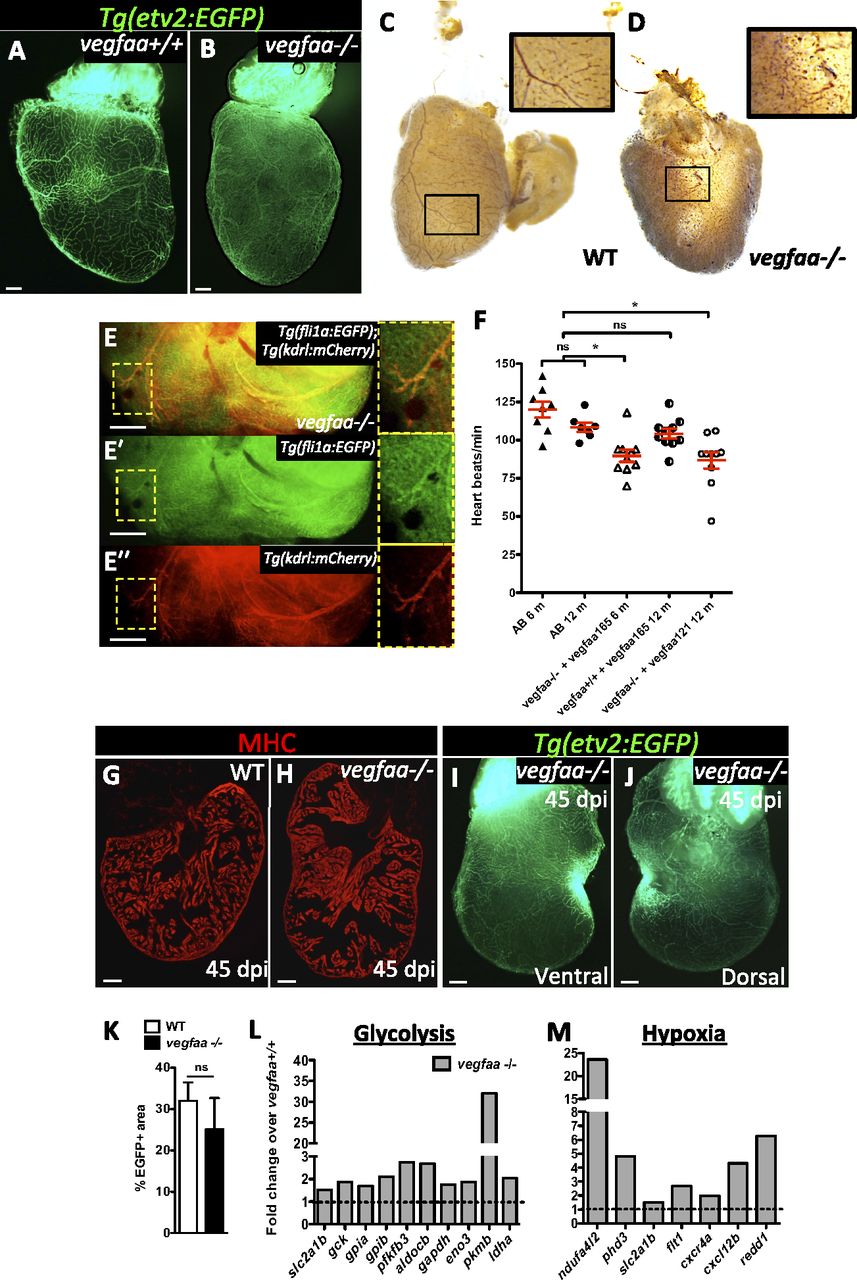
vegfaa-/- hearts exhibit features of myocardial hibernation and can regenerate. (A and B) Ventral views of Tg(etv2:EGFP) vegfaa+/+ and vegfaa-/- adult hearts. (C and D) Erythrocytes in WT and vegfaa-/- hearts stained with O-dianisidine to assess vascular integrity. Insets show high-magnification images of coronaries containing erythrocytes. (E-E′′) Dorsal view of Tg(fli1a:EGFP);Tg(kdrl:mCherry) vegfaa-/- ventricle. Insets show fli1a:EGFP+ and kdrl:mCherry+ coronaries. (F) Doppler measurements of heart rate in WT fish (AB) of 6 (n = 8) and 12 (n = 8) months of age, 6-mo-old vegfaa-/- (n = 10) and 12-mo-old vegfaa+/+ (n = 10) injected with 200 pg of vegfaa 165, and 12-mo-old vegfaa-/- (n = 10) injected with 50 pg of vegfaa 121. Each data point represents a single fish. Mean ± SEM is indicated in red. ns, no significant differences. *P < 0.05. (G and H) Sections of WT (n = 4) and vegfaa-/- (n = 4) hearts at 45 dpi. Cardiomyocytes are immunostained with anti-MHC antibody (red). (I and J) Ventral and dorsal views of a Tg(etv2:EGFP) vegfaa-/- heart at 45 dpi. (K) Percentage of etv2:EGFP+ area in the apex of WT (n = 5) and Tg(hsp70l:dn-vegfaa) ventricles (n = 3) at 45 dpi. (L and M) Differentially expressed genes involved in glycolysis or stimulated by hypoxia obtained by microarray profiling of vegfaa+/+ and vegfaa-/- adult ventricles. Data are expressed relative to vegfaa+/+ ventricles (dashed lines). (Scale bars: 100 µm.)

