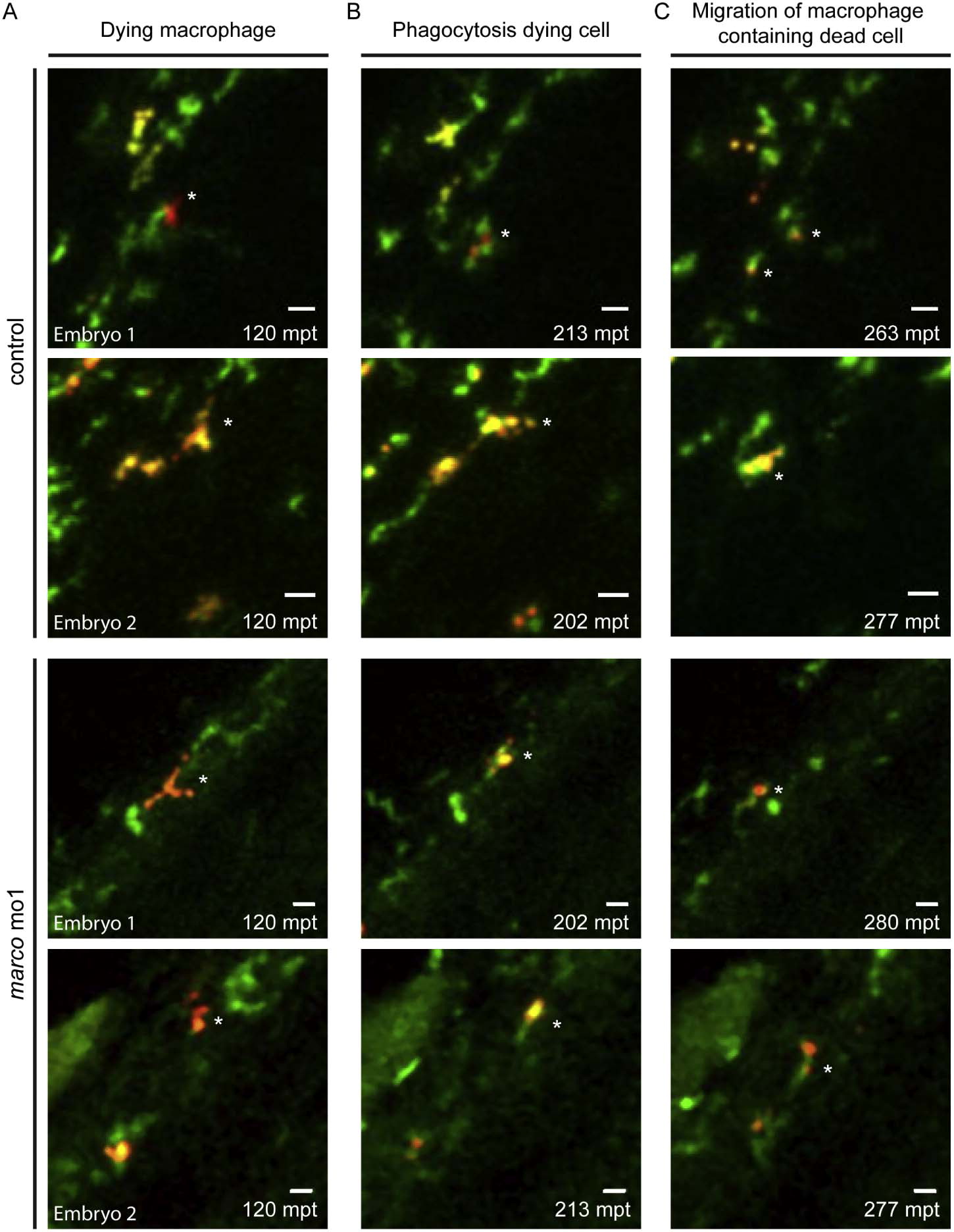
Fig. 3
Morphants of marco are capable of phagocytosing dead cells. Phagocytosis of dying cells by macrophages was monitored in time. marco morphants and their controls were imaged after metronidazole treatment which induces nitroreductase-mediated cell ablation in a subpopulation of macrophages (mCherry positive cells). All macrophages express GFP. Time points represented are (A) time of nitroreductase-positive cells undergoing cell death, (B) phagocytosis of the dying cells by healthy GFP-positive macrophages and (C) migration of the macrophage containing the dead cell. Time lapse recordings were made of 4 embryos per group and examples of phagocytosis are shown for 2 control embryos en 2 marco morphants. Quantification of phagocytosis events are shown in Supplementary Fig. S2B. Asterisks indicate the phagocytosing macrophage; mpt: minutes post treatment.

