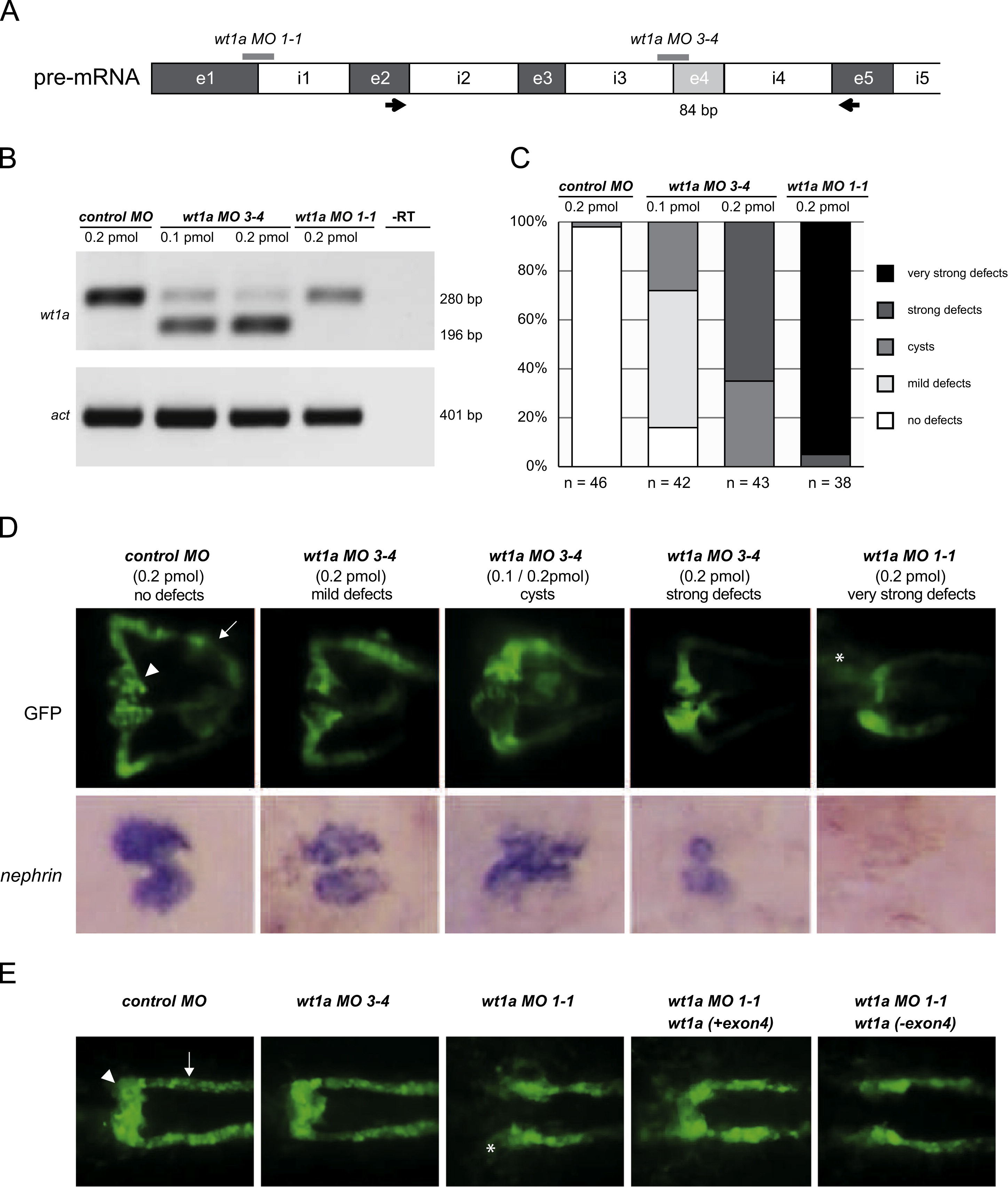
Fig. 4
Knockdown of wt1a exon4 in zebrafish. To assess the in vivo function of wt1a exon4 sequences we employed a morpholino-based knockdown approach in zebrafish. (A) Scheme of wt1a pre-mRNA with intron-exon structure. Thick gray lines indicate the target sites of the morpholinos. Arrows represent the location of the primer pair used to analyze the knockdown efficiency by RT-PCR. (B) Assessment of the knockdown efficiency of wt1a splice morpholinos. RT-PCR analysis was performed from total RNA of 10 pooled embryos 48 h after morpholino injection using the primer pair indicated in (A). Both morpholinos cause efficient reduction of correctly spliced mRNA represented by the band at 280 nucleotides. The lower band corresponds to wt1a mRNA with exon 4 deletion triggered by the wt1a MO3-4 morpholino. The wt1a MO1-1 morpholino causes insertion of intron1. The product, however, is not amplified under the PCR conditions. (C) Statistical analysis of embryonic kidney (pronephros) defects following the injection of wt1a morpholinos. Malformations were grouped into different categories. (D) Morpholino effects on pronephros development and nephrin expression. All embryos were imaged at 48 hpf and are shown in dorsal view with anterior to the left. Upper row: Representative images of normal and malformed embryonic kidneys of transgenic wt1b::GFP zebrafish. Normally developed kidneys consist of two nephrons with fused glomeruli (arrowhead) and pronephric tubules (arrow). In wt1a splice 3-4 morphants three categories of defects were observed: less differentiated and often not fused glomeruli (mild defects), symmetric or asymmetric cysts (cysts) and completely undifferentiated glomeruli (strong defects). Splice 1-1 morphants failed to form glomeruli and a significant number of GFP positive cells were found outside of the pronephric field (asterisk, very strong defects). Bottom row: Micrographs of whole mount in situ hybridization with the podocyte marker nephrin. Injection of the wt1a MO3-4 morpholino results in significant expression changes compared to control-injected embryos: a less intensive and more diffuse expression was detected in the group of embryos with mild defects. Embryos with cysts displayed a stretched and stripe-like expression pattern whereas a significant reduction of expression was detected in the group of embryos with strong defects. Wt1a MO1-1 injection causes a complete loss of expression. (E) Wt1a(+exon 4) mRNA partially rescues the kidney defects in wt1a morphant embryos. All embryos were imaged at 24 hpf and are shown in dorsal view with anterior to the left. Normal patterning of the pronephros in the transgenic wt1b::GFP line gives rise to fusing pronephric glomeruli (arrowhead). Tubules are also clearly visible and marked by an arrow. In wt1a splice 3-4 morphants, pronephric development is comparable to control with no obvious defects. In contrast, splice 1-1 morphants fail to develop glomeruli and a significant number of GFP-positive cells can be found outside of the pronephric field (asterisk). Injection of mRNA encoding wt1a(+exon 4) but not wt1a(exon 4) partially rescues this drastic impairment of pronephros development.
Reprinted from Developmental Biology, 393(1), Schnerwitzki, D., Perner, B., Hoppe, B., Pietsch, S., Mehringer, R., Hänel, F., Englert, C., Alternative splicing of Wilms tumor suppressor 1 (Wt1) exon 4 results in protein isoforms with different functions, 24-32, Copyright (2014) with permission from Elsevier. Full text @ Dev. Biol.

