
|
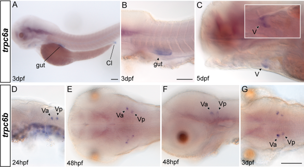
Fig. 7
A-G: Expression pattern of trpc6a (A-C) and trpc6b (D-G). trpc6a expression is not detectable before 3 days postfertilization (dpf). A: Lateral overview of WISH in a larva 3 dpf. B: Expression of trpc6a in the gut is shown in a lateral view. C: The heart ventricle (V) expresses the gene, too. Insert is a ventral view on the heart to compare with the lateral view in the main image. D: Expression of trpc6b is present in the hindbrain region 24 hr postfertilization (hpf), lateral view. E,F: Expression of trpc6b in embryos 48 hpf shown in dorsal (E) and lateral (F) views. G: Dorsal head view on the expression in larvae 3 dpf. Anterior is always left. Cl, cloaca; Va, anterior clusters of trigeminal motor neurons; Vp, posterior clusters of trigeminal motor neurons. Scale bar = 100 µm in A (for all pictures of not otherwise indicated), B,G.