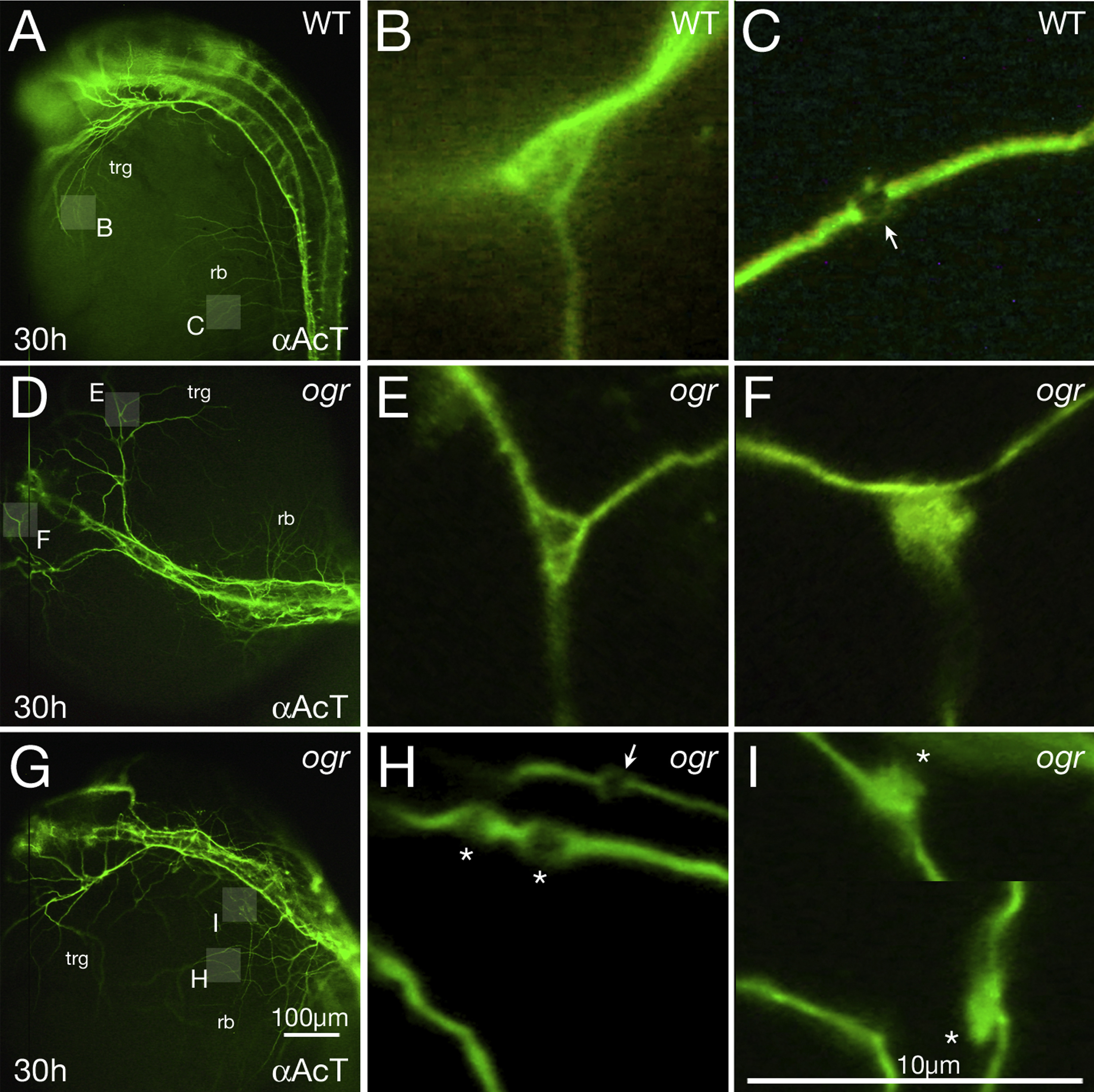
Fig. 8
Microtubules are disorganized in mutant axons. (A-I) 30 h embryos visualized with anti-Acetylated Tubulin. (A, D, G) Low magnification, showing pathfinding errors in trigeminal (trg) and Rohon-Beard (rb) peripheral axons; highlighted boxes indicate regions shown at higher magnification in panels (B, C, E, F, H, I). (B, C) Wild type: (B) a branch point where individual microtubules defasciculate and fan out and (C) a presumptive presynaptic swelling (arrow) where individual microtubules form a loop. (E, F) ogre: (E) a normal looking branch point and (F) a branch point where microtubules tangle. (H, I) ogre: (H) three presumptive presynaptic swellings where one looks normal (arrow) and two not normal (indicated with asterisk) due to them being immediately adjacent to each other and appearing swollen (the one on the left may also be instead a region where individual microtubules abnormally defasciculate from one another and tangle), and (I) two presumptive presynaptic swellings along the same neurite (the picture is cropped to show both) where instead of forming loops the microtubules form large irregular tangles (asterisks). Note how in general mutant presumptive presynaptic swellings are larger than wild type.
Reprinted from Developmental Biology, 418(2), Warga, R.M., Wicklund, A., Richards, S.E., Kane, D.A., Progressive loss of RacGAP1/ogre activity has sequential effects on cytokinesis and zebrafish development, 307-22, Copyright (2016) with permission from Elsevier. Full text @ Dev. Biol.

