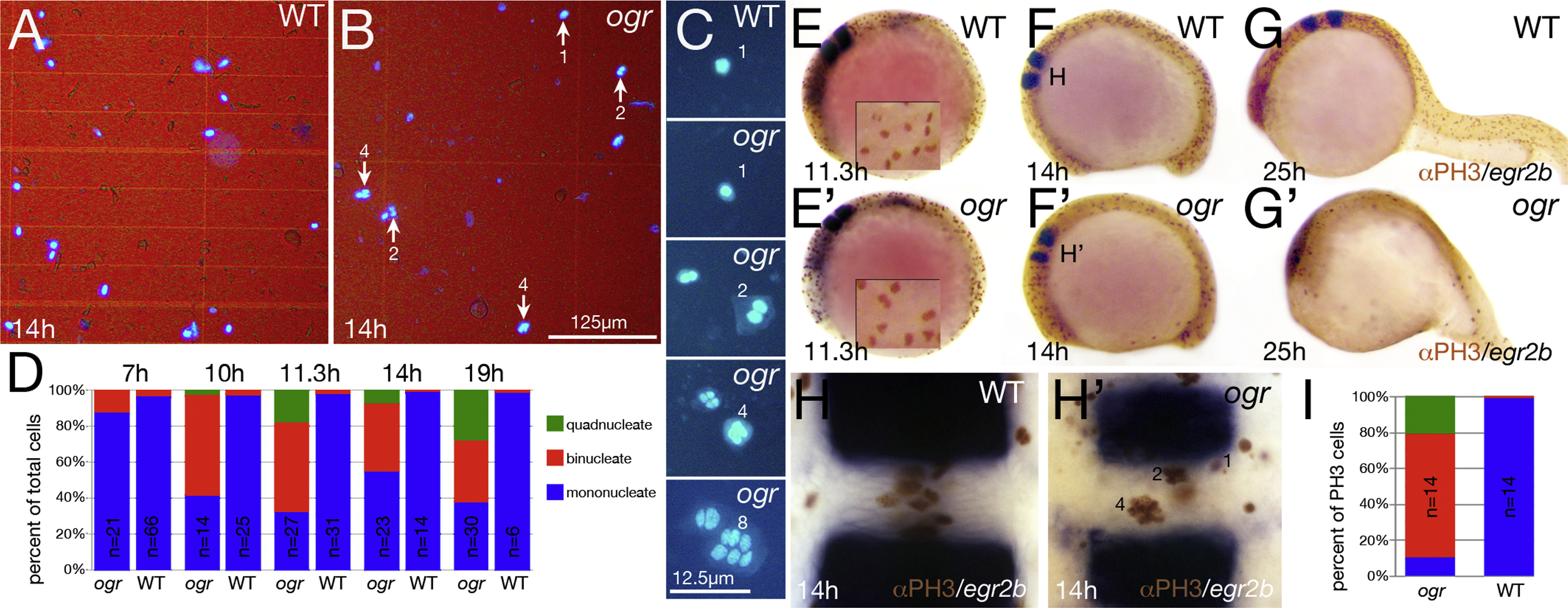
Fig. 4
Mitosis continues after cytokinesis fails. (A-D) Dissociated cells. (A, B) Low magnification and (C) high magnification showing range of nuclear phenotypes. (D) Quantification of multinucleate cells. At 7 h, the proportion of cells with double nuclei ranked embryos; mutant embryos were then inferred by the simple Mendelian segregation of embryos with the highest deviation from wild type patterns. At 10 and 11.3 h, mutant embryos were inferred based on cell counts deviating from the wild type proportions. At 14 and 19 h, mutant embryos were identified morphologically before cell dissociations. The results of all of the embryos along with a partial statistical analysis are in Supplemental Fig. 2. (E-I) Multinucleate cells are mitotically active. (E-H) Phospho-Histone H3 staining and egr2b expression to visualize rhombomere 4, (E-G) are side views, insets in (E) are higher magnification of mitotic nuclei and (H) high magnification dorsal view of rhombomere 4. (I) Quantification of mitotic multinucleate cells at 10-somites in rhombomere 4; n indicates the number of embryos scored. Data are significantly different at the p<<0.001 level (contingency table tested with Pearson′s chi square).
Reprinted from Developmental Biology, 418(2), Warga, R.M., Wicklund, A., Richards, S.E., Kane, D.A., Progressive loss of RacGAP1/ogre activity has sequential effects on cytokinesis and zebrafish development, 307-22, Copyright (2016) with permission from Elsevier. Full text @ Dev. Biol.

