
|
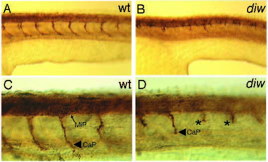
Fig. 8
Axonal outgrowth of primary motorneurons are affected in diw mutants. Antibody labeling with the znp-1 antibody stains axonal projections of primary motorneurons in 30-hour wild type (A,C) and residual axons in diw mutant embryos (B,D). (A) Lateral view of a wild-type embryo stained with znp-1. Per segment, one CaP axon is visible. In diw mutants these axons are shorter (B). (C) Higher magnification of the same wild-type embryo as in A. The MiP and the CaP axons are labeled. (D) Higher magnification of the same diw embryo as in B. Short, CaP-like axons are stained (asterisk). No axons resembling dorsally projecting MiP axons are labeled.