
|
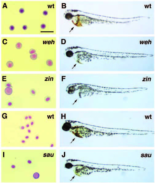
Fig. 3
Mutations in the genes weißherbst (weh), zinfandel (zin) and sauternes (sau) result in embryos with hypochromic blood and decreasing blood cell counts. (A,C,E,G,I) Isolated blood cells from wild-type and mutant larvae stained by the Wright-Giemsa method. (B,D,F,H,J) O-dianisidine staining for hemoglobin (arrows) in wildtype and mutant embryos. (A) Blood cells from 2-day old wild-type embryos have the normal morphology of late erythroblasts. (B) Wild type embryos express large amounts of hemoglobin in their differentiating red blood cells. (C) Blood cells from 2-day old wehtp85c mutant embryos are larger with open nuclei more typical of early proerythroblasts. (D) 2-day old wehtp85c mutant embryos have approximately 50% of the normal number of blood cells which express much lower levels of hemoglobin compared to wild-type blood cells. (E) Blood cells of 2-day old zinte207 mutant embryos are large but are more differentiated than the blood cells of weh mutant embryos. (F) 2-day old zinte207 heterozygotes (shown here) and homozygotes both lack detectable hemoglobin. (G) 3-day old wildtype embryos have mature erythrocytes with condensed nuclei and red staining hemoglobin. (H) A wild-type, 3-day old, embryo has large amounts of blood expressing hemoglobin. (I) Blood cells of 3- day old sauy121a mutant embryos are relatively normal in morphology, but they are reduced in number to between 25% and 50% of normal levels with a few large basophilic cells. (J) 3-day old sauy121a mutant embryos have fewer blood cells and thus a reduced total amount of hemoglobin expression. The scale bar in panel (A) is equal to 20 microns for panels (A,C,E,G,I).