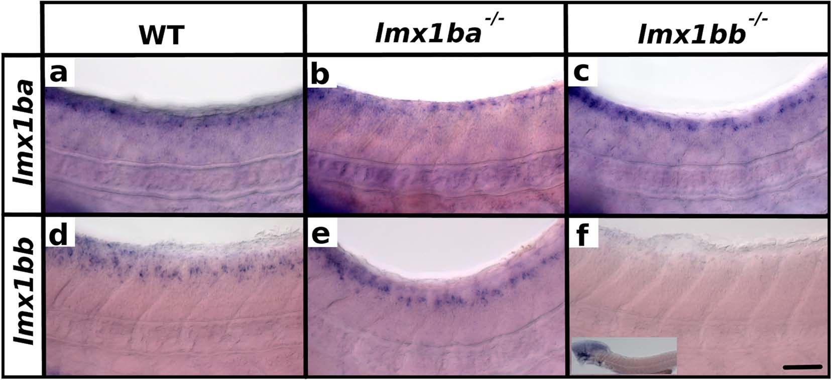
Fig. 2
Expression of lmx1b RNAs in lmx1b mutants. Lateral view of zebrafish spinal cord at 48 h (a-f). Anterior left, dorsal top. in situ hybridization of lmx1ba (a-c) or lmx1bb (d-f) in WT (a and d), lmx1ba mutant (b and e) and lmx1bb mutant (c and f). Lower magnification insert in (f) shows expression remaining in hindbrain region. The rest of the head was removed for genotyping. One in situ hybridization of at least 40 embryos was conducted for each of b and e. Two independent in situ hybridizations of at least 50 embryos each were conducted for a, c, d and f. In these cases, results were the same for each replicate experiment. At least three genotyped mutant and wild-type embryos were analyzed in detail for each experiment. Scale bar = 50 µm

