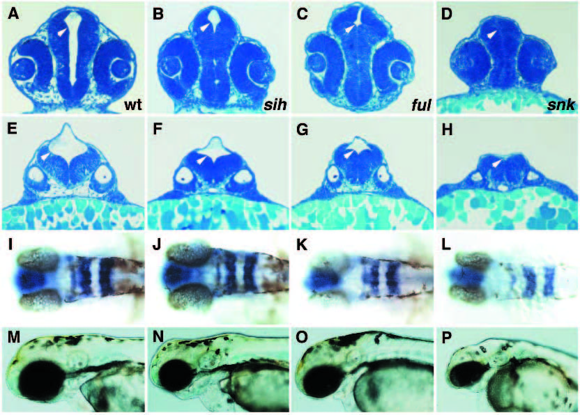
Fig. 8
Phenotypic analysis of mutations affecting ventricle enlargement. (A,E,I,M) Wild type; (B,F,J,N) silent heart (sih)b109; (C,G,K,O) fullbrain (ful)m133; (D,H,L,P) snakehead (snk)m273. (A,B,C,D) Transverse sections through eye and lens at 28 hpf. (E,F,G,H) Transverse section through anterior ear and otolith at 28 hpf. Note the different degrees of ventricle (white arrowheads) reduction in sih, ful and snk. (I,J,K,L) Expression of rtk1 in rhombomeres 1,3 and 5 at 31 hpf; dorsal view. (M,N,O,P) Wild-type and mutant embryos at 53 hpf. Note the differences in reduction in the size of the brain and the onset of degeneration in sih, ful and snk.

