Image
Figure Caption
Fig. S5
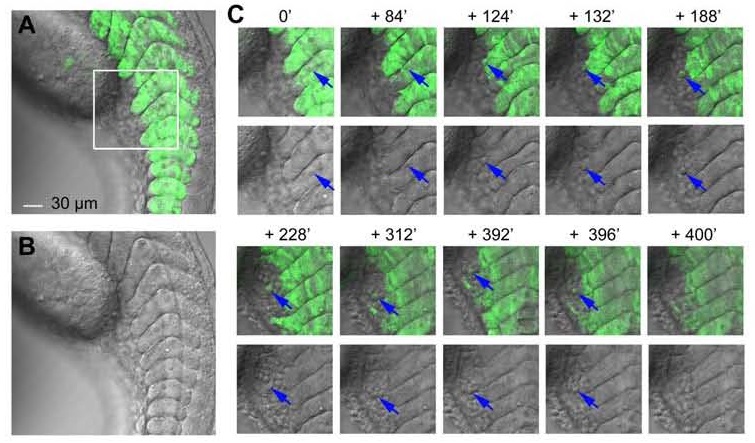
Emigration of an EOS-positive cell from the ventral domain of the somite in Tg(foxc1b:EOS) transgenic embryo. The embryo was laterally orientated and observed continuously from 22 to 30 hpf with focus on posterior somites by confocal microscopy. The imaging time points were indicated. An emigrating cell was indicated by arrows. See also Movie S3. Note that the embryo looked younger because its posterior trunk could not extend well when it was embedded in agarose gel. Beside, the tracked cell was vanished in the last frame most likely because it had flowed away with the circulation.
Acknowledgments
This image is the copyrighted work of the attributed author or publisher, and
ZFIN has permission only to display this image to its users.
Additional permissions should be obtained from the applicable author or publisher of the image.
Full text @ J. Mol. Cell Biol.

