
|
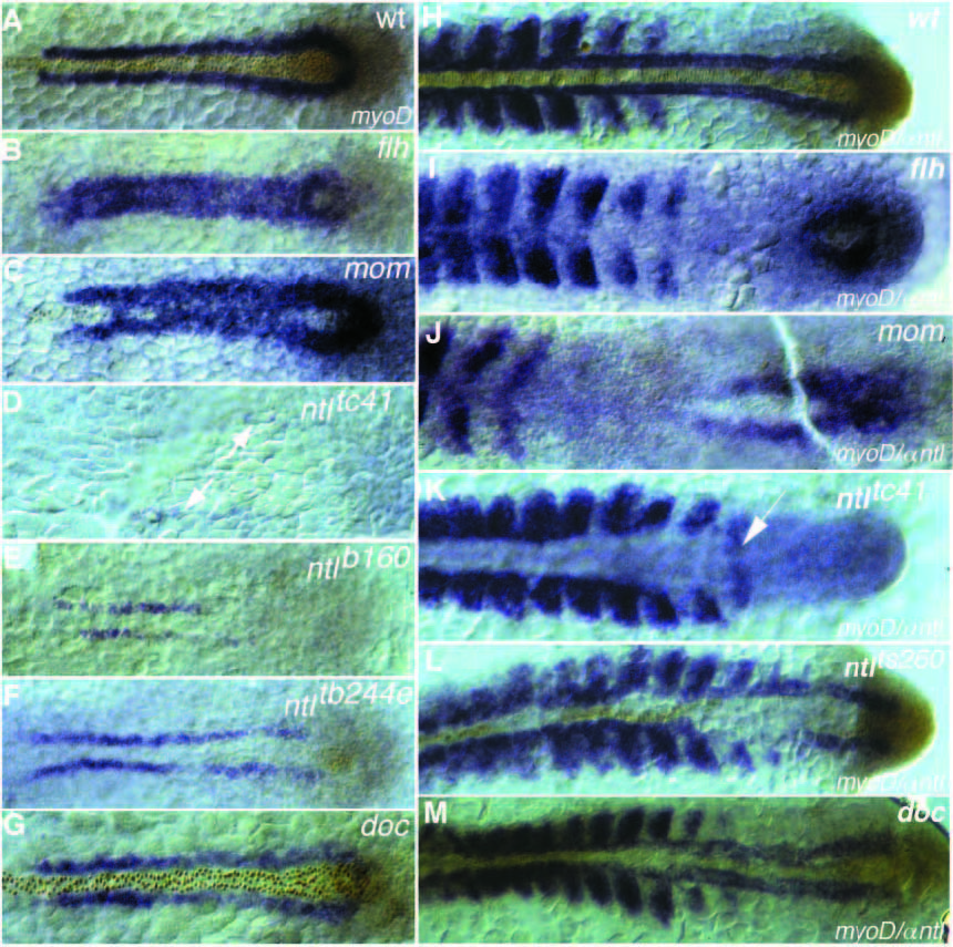
Fig. 6
Dorsal view of myoD RNA (blue staining) and Ntl antibody staining (brown nuclear staining) at (A-G) tailbud stage and (H-M) 15- somite stage of (A,H) wild-type, (B,I) flhtk241, (C,J) momth211, (D,K) ntltc41, (E) ntlb160, (F) ntltb244e, (L) ntlts260 and (G,M) doctt258 embryos. (A) In wild type at the tailbud stage myoD expression is restricted to a line of cells left and right of the notochord. (B,C) The gap between the two stripes is filled with myoD expressing cells in (B) flhtk241 and (C) momth211 embryos. ntl expression is detectable only in the tailbud and sometimes in a few cells anteriorly, which do not express myoD (C). (D) Strongly reduced myoD expression a greater distance from the midline is detectable in embryos with the strong allele, ntltc41(arrows). myoD expression at the 15-somite stage in (H) wild type is expressed in the adaxial cells and in the posterior part of the somites. In (I) flhtk241 embryos and (J) momth211 embryos myoD expression in the adaxial cells is not maintained. (K) In embryos with the strong allele, ntltc41, somites are separated by a big gap of nonexpressing cells, whereas the 15th somite is fused in the midline (arrow). (L) In embryos with the weak allele, ntlts260, adaxial cells next to ntl-expressing cells express myoD at a higher level than the somites on the opposite side. (M) In doctt258 embryos adaxial expression in anterior regions is reduced and closer together.