
|
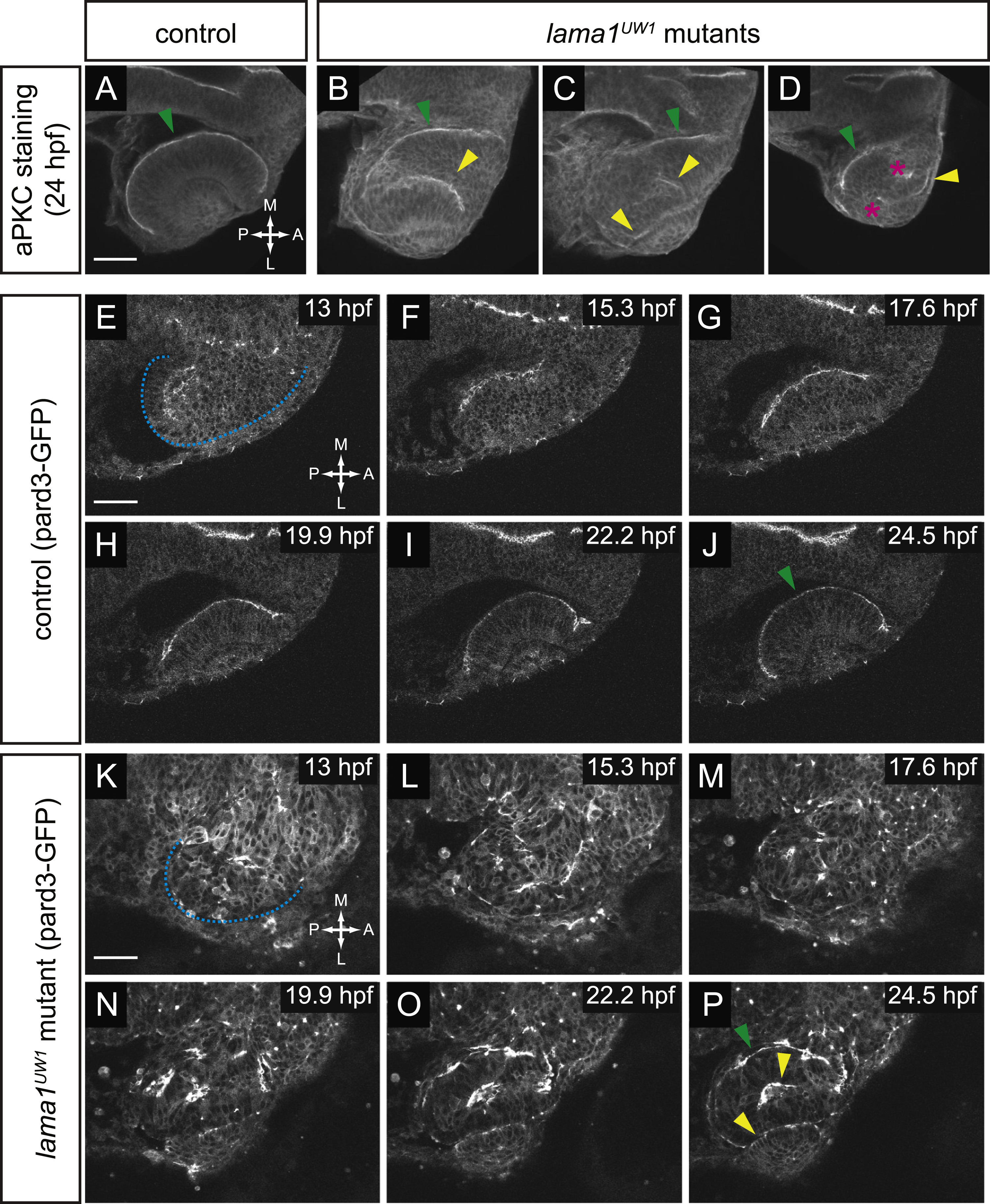
Fig. 6
Apicobasal polarity is disrupted from the earliest stages of optic vesicle morphogenesis. (A-D) Antibody staining for aPKC in control (A) and lama1UW1 mutant embryos (B-D) reveals disruption of polarity at 24 hpf. green arrowheads, correct apical domain. yellow arrowheads, ectopic apical domains. asterisks, ectopic puncta. (E-P) Single confocal sections from 4D datasets of apical domain dynamics (marked by pard3-GFP) in a lama1UW1 control embryo (E-J), or a mutant embryo (K-P). In lama1UW1 mutant embryos, pard3-GFP localization is disrupted similar to aPKC. dashed blue line marks outline of optic vesicle. green arrowheads, correct apical domain. yellow arrowheads, ectopic apical domains. Dorsal views; scale bar, 50 µm. A, anterior; P, posterior; M, medial; L, lateral.
Reprinted from Developmental Biology, 416(2), Bryan, C.D., Chien, C.B., Kwan, K.M., Loss of laminin alpha 1 results in multiple structural defects and divergent effects on adhesion during vertebrate optic cup morphogenesis, 324-37, Copyright (2016) with permission from Elsevier. Full text @ Dev. Biol.