Fig. S5
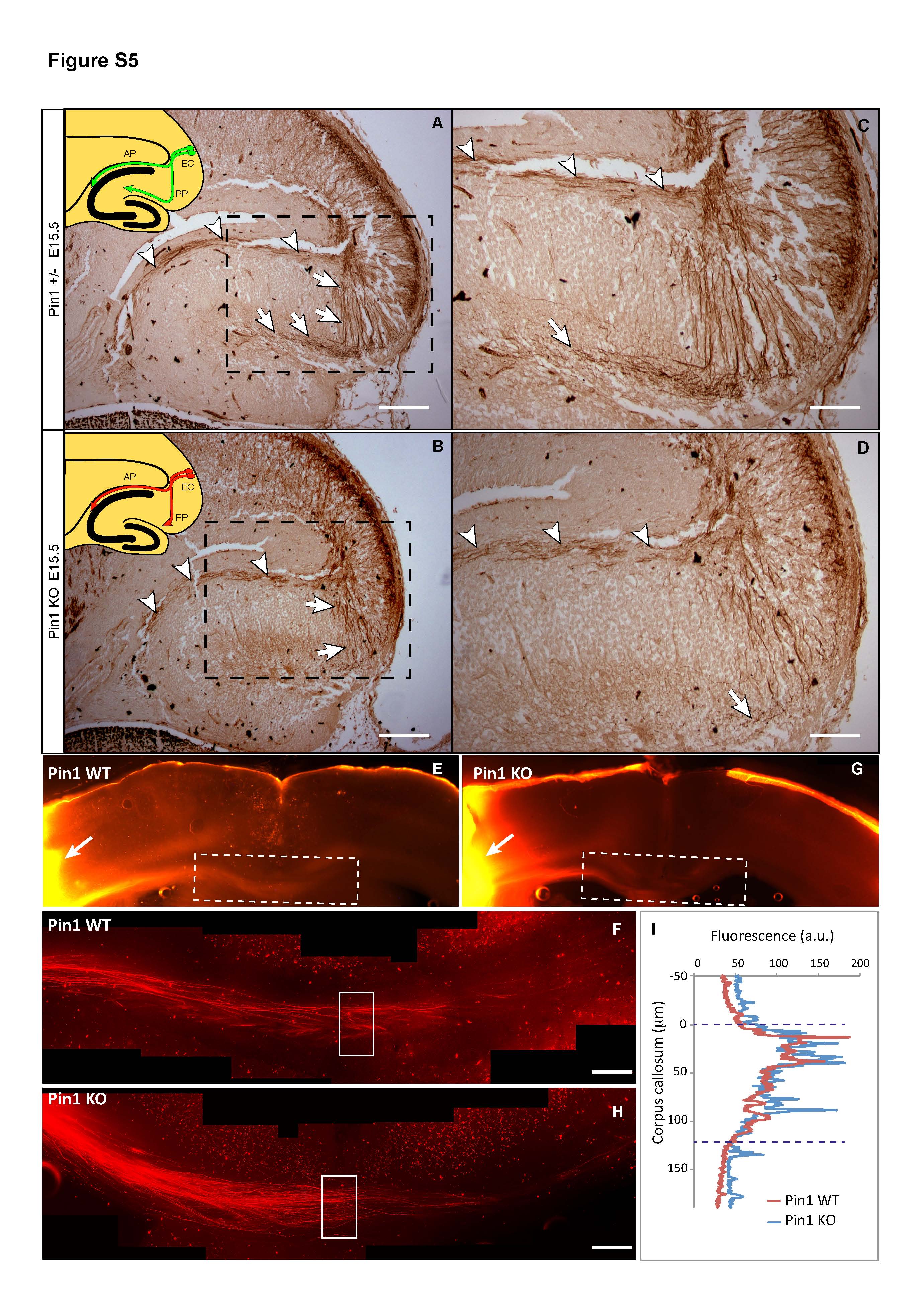
Alvear pathway of the enthorino-hippocampal projections at E15.5 Pin1 KO embryos and fasciculation of somatosensory (S1) axons at midline of corpus callosum in adult Pin1 KO mice. Related to Figure 6
NF-L immunostaining of E15.5 brain horizontal sections shows normal development of the Pin1 KO (B, D) alvear projections (arrowheads), while growth of perforant fibers is significantly reduced (arrows) when compared to Pin1 WT littermates (A, C). The fasciculation of S1 axons in midline corpus callosum is similar in Pin1 WT (E, F) and Pin1 KO (G, H) adult mice (3 month old) (arrows indicates the place of DiI crystal insertion). (I) Fluorescence intensity of S1 axons along the D-V axis at the midline shows similar distribution and diameter of the axon bundle in Pin1 WT and Pin1 KO mice. EC-entorhinal cortex, PP-perforant pathway, AP-alvear pathway (Scale bars: A, B 200 µm; C, D, F, H 100 µm). (F, H are composite images of 6 and 7 images, respectively).

