Fig. 4
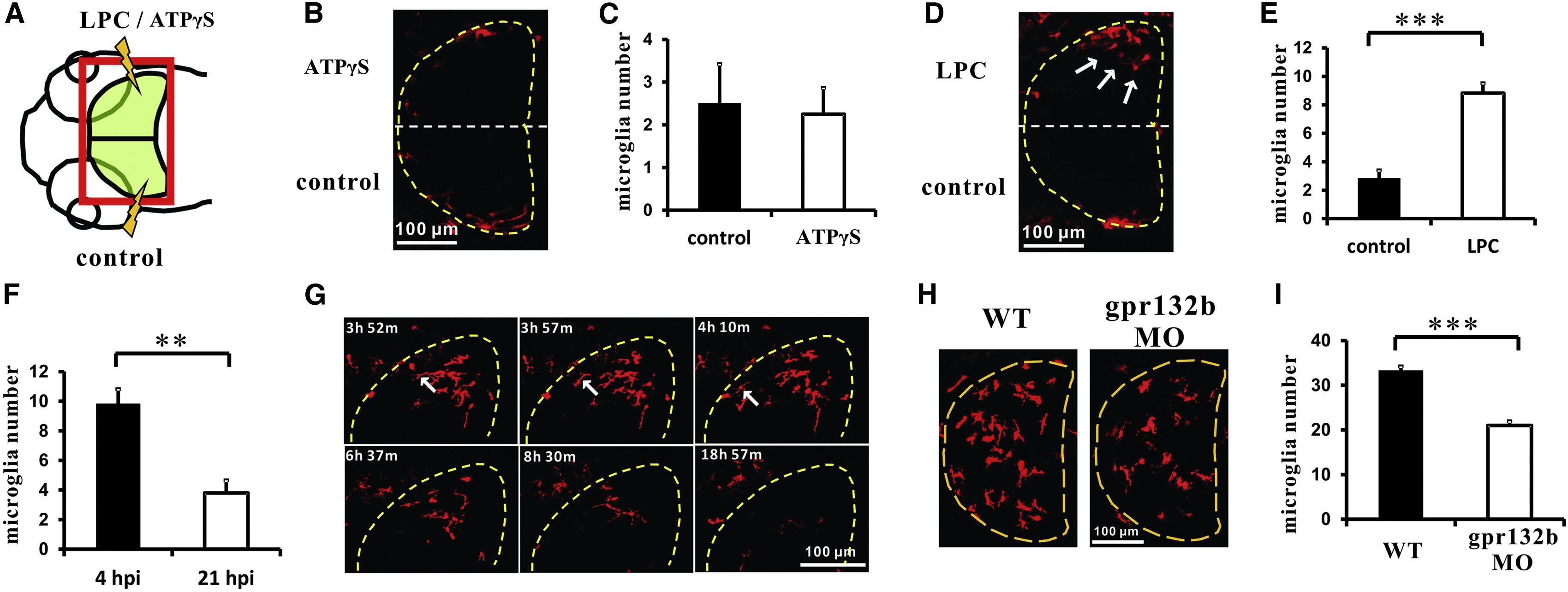
LPC Promotes the Entry of Microglial Precursors into the Brain
(A) A schematic diagram of the dorsal view of zebrafish head. The red square indicates the region where LPC or ATPγS is injected. Normally the upper half-brain and lower half-brain are injected with LPC or ATPγS and control buffer, respectively.
(B) Dorsal view of the ATPγS-injected brain of Tg(Xla.Tubb:bcl-2;mpeg1:loxP-DsRedx-loxP-GFP) embryos at 4-5 hpi. The upper half-brain is injected with ATPγS and the lower half-brain is injected with control buffer. DsRedx+ cells represent microglia. The midbrain is labeled by dashed lines.
(C) Quantification of microglia in ATPγS-injected brain of Tg(Xla.Tubb:bcl-2;mpeg1:loxP-DsRedx-loxP-GFP) embryos at 4-5 hpi. n = 8 for control and ATPγS injection. DsRedx+ cells represent microglia. Error bars represent the mean ± SEM.
(D) Dorsal view of the LPC-injected brain of Tg(Xla.Tubb:bcl-2;mpeg1:loxP-DsRedx-loxP-GFP) embryos at 4-5 hpi. The upper half-brain is injected with LPC and the lower half-brain is injected with control buffer. DsRedx+ cells represent microglia. White arrows indicate LPC-induced microglia in the brain. The midbrain is labeled by dashed lines.
(E) Quantification of microglia in the LPC-injected brain of Tg(Xla.Tubb:bcl-2;mpeg1:loxP-DsRedx-loxP-GFP) embryos at 4-5 hpi. n = 17 for control and LPC injection. Error bars represent the mean ± SEM. ***p < 0.001.
(F) Quantification shows that the number of microglia in the LPC-injected brain of Tg(Xla.Tubb:bcl-2;mpeg1:loxP-DsRedx-loxP-GFP) embryos is drastically decreased by 21 hpi. DsRedx+ cells represent microglia. n = 5. Error bars represent the mean ± SEM. **p < 0.01.
(G) Time-lapse imaging pictures show that the microglia in the LPC-injected brain of Tg(mpeg1:loxP-DsRedx-loxP-eGFP) embryos gradually migrate out of the brain. DsRedx+ cells represent microglia. White arrowheads indicate one microglia migrating out of the brain. Dashed lines indicate the midbrain.
(H) Dorsal view images of the midbrain of Tg(mpeg1:loxP-DsRedx-loxP-eGFP) control and gpr132b morphants (MO). DsRedx+ cells represent microglia. Dashed lines indicate the midbrain.
(I) Quantification of the number of microglia number in the midbrain of Tg(mpeg1:loxP-DsRedx-loxP-eGFP) control and gpr132b morphants (MO). n = 14 for WT control and n = 15 for MO. Error bars represent the mean ± SEM. ***p < 0.001.
See also Figure S4 and Movie S4.
Reprinted from Developmental Cell, 38(2), Xu, J., Wang, T., Wu, Y., Jin, W., Wen, Z., Microglia Colonization of Developing Zebrafish Midbrain Is Promoted by Apoptotic Neuron and Lysophosphatidylcholine, 214-22, Copyright (2016) with permission from Elsevier. Full text @ Dev. Cell

