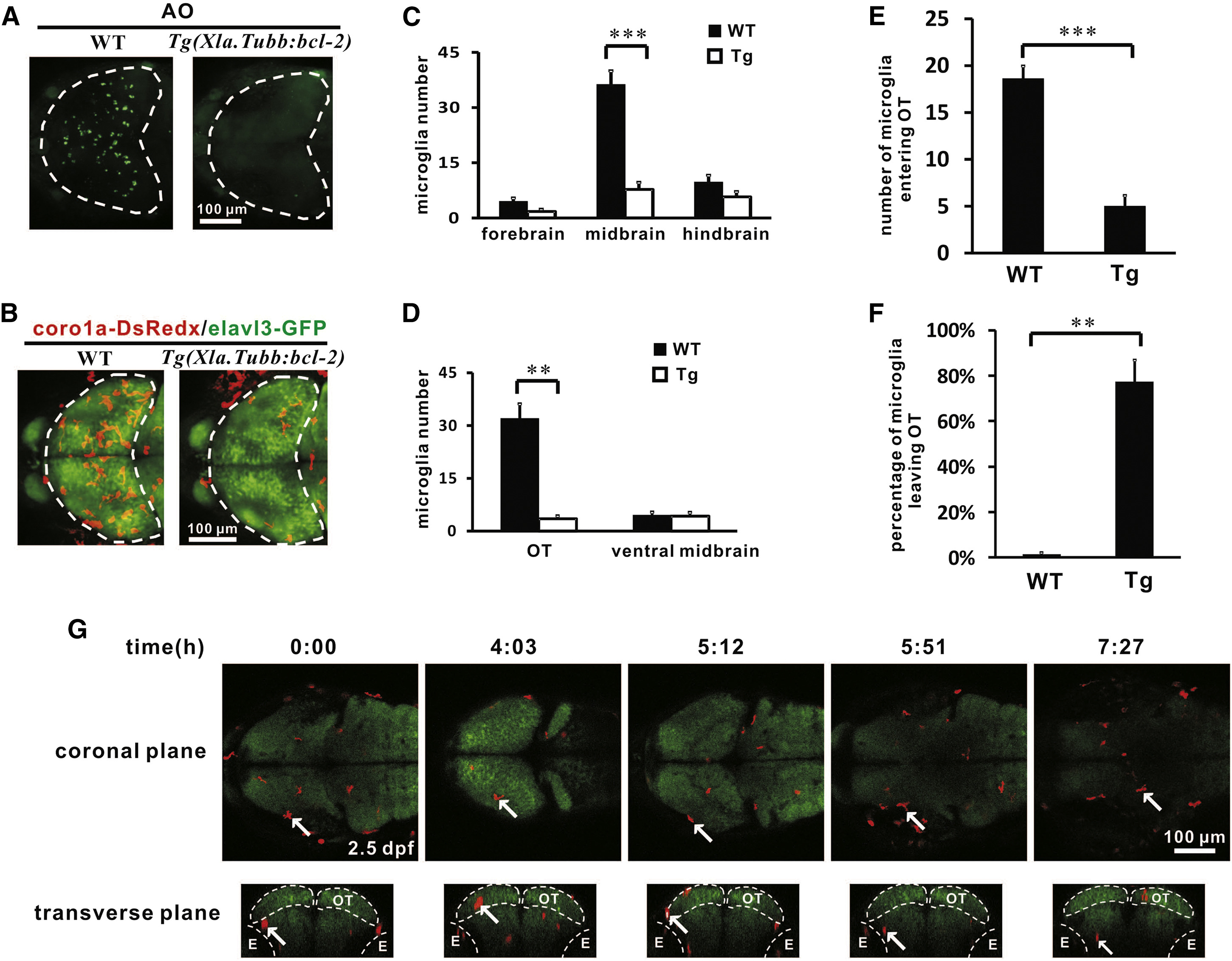
Fig. 3
Neuron-Specific bcl-2 Overexpression Suppresses Neuronal Cell Death and Blocks Microglia Colonization of the Optic Tectum
(A) Acridine orange (AO) staining reveals that neuronal cell death is largely prevented in the optic tectum of 3 dpf Tg(Xla.Tubb:bcl-2) embryos. The optic tectum is indicated by dashed lines.
(B) The number of microglia in the optic tectum is drastically reduced in the optic tectum of 3 dpf Tg(Xla.Tubb:bcl-2) embryos. The optic tectum is indicated by dashed lines. Red and green signals represent coro1a-DsRedx+ microglia and elavl3-GFP+ neurons, respectively.
(C) Quantification of the number of microglia in the forebrain, midbrain, and hindbrain of 3 dpf WT and Tg(Xla.Tubb:bcl-2) embryos. Error bars represent the mean ± SEM. ***p < 0.001 (n = 4).
(D) Quantification of the number of microglia in the optic tectum and ventral midbrain of 3 dpf WT and Tg(Xla.Tubb:bcl-2) embryos. Error bars represent the mean ± SEM. **p < 0.01 (n = 4).
(E) Quantification of the number of microglia that migrate into the optic tectum within the 24 hr imaging period (from 2 dpf to 3 dpf) of WT and Tg(Xla.Tubb:bcl-2) embryos. Error bars represent the mean ± SEM. ***p < 0.001 (n = 5 for WT embryos, n = 4 for transgenic embryos).
(F) Quantification of the percentage of microglia that shuffle out of the optic tectum within the 24 hr imaging period (from 2 dpf to 3 dpf) of WT and Tg(Xla.Tubb:bcl-2) embryos. Error bars represent the mean ± SEM. **p < 0.01 (n = 5 for WT embryos, n = 4 for transgenic embryos).
(G) Coronal (top) and transverse (bottom) views of time-lapse imaging pictures of the midbrain show a typical microglia (labeled by the white arrow) that enters the optic tectum and subsequently migrates out of the optic tectum in the Tg(Xla.Tubb:bcl-2) embryos. The optic tectum and eyes are indicated by dashed lines. Red and green signals represent coro1a-DsRedx+ microglia and elavl3-GFP+ neurons, respectively. E, eye; OT, optic tectum.
See also Figure S3 and Movie S3.
Reprinted from Developmental Cell, 38(2), Xu, J., Wang, T., Wu, Y., Jin, W., Wen, Z., Microglia Colonization of Developing Zebrafish Midbrain Is Promoted by Apoptotic Neuron and Lysophosphatidylcholine, 214-22, Copyright (2016) with permission from Elsevier. Full text @ Dev. Cell

