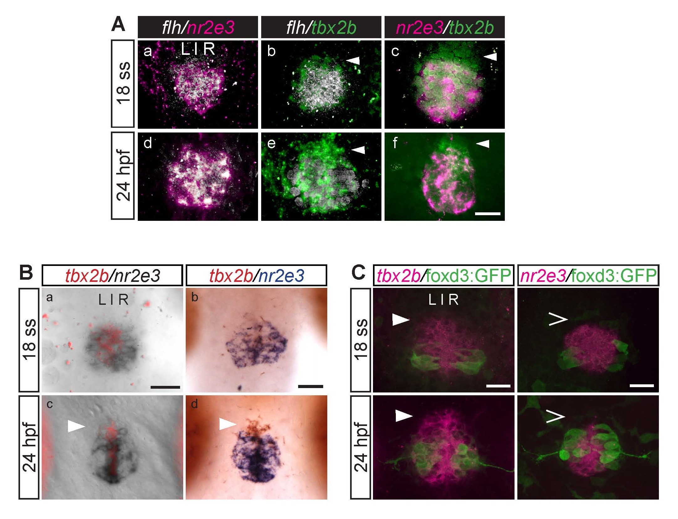
Fig. S5
During parapineal development, the anterior-most region of the pineal anlage is mostly tbx2b-positive, nr2e3- and flh-negative (arrowheads). This difference is subtle at the 18 ss (18 hpf) but becomes more apparent at 24 hpf. (A) Dorsal views of the pineal anlage of double fluorescence in situ hybridizations of flh/nr2e3, flh/tbx2b, and nr2e3/tbx2b embryos at 18 ss (a-c) and 24 hpf (d-f). nr2e3 and flh expression appear to mostly overlap (a, d). (B) Double in situ hybridizations of tbx2b and nr2e3 in the pineal anlage at the indicated stages. (a,c) tbx2b was developed using Fast Red reagents and imaged under fluorescence; nr2e3 was developed using NBT/BCIP and imaged under bright field. (b,d) tbx2b (developed using INT/BCIP) and nr2e3 (developed using NBT/BCIP) were imaged under bright field. (C) Double labeling with fluorescence in situ hybridization (tbx2b or nr2e3) and antibody labeling of foxd3:GFP positive cells (used as landmarks) in the pineal anlage. At these stages, foxd3:GFP is only expressed in projection neurons. Scale bars: 30 µm.

