Image
Figure Caption
Fig. S4
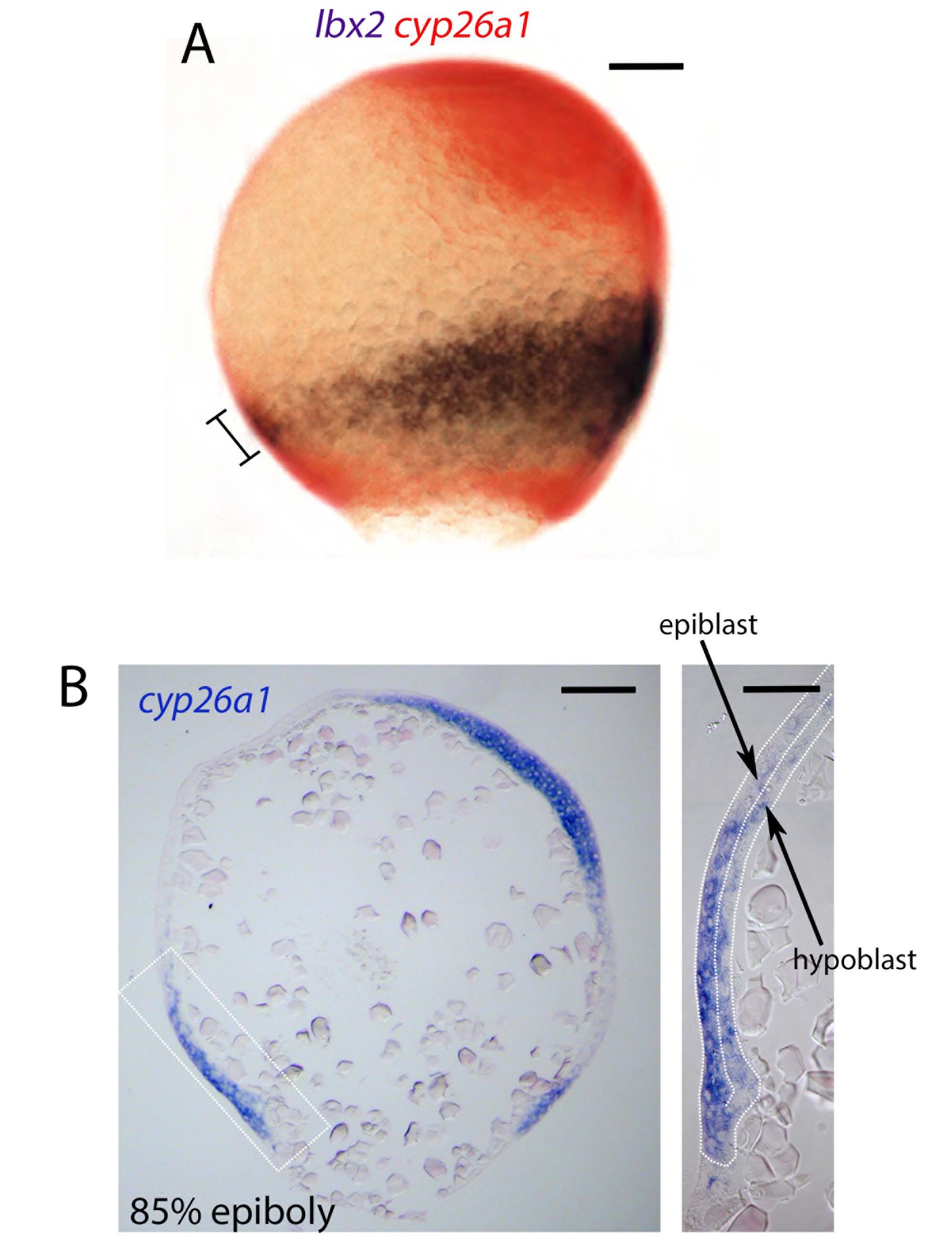
Expression of cyp26a1 and lbx2.
A) Detection of Ibx2 (purple/black) and cyp26a1 (red) transcripts by whole mount in situ hybridization is shown at the 85% epiboly stage. The embryo is viewed laterally with the dorsal midline on the right. Bar indicates overlap between cyp26a1 and Ibx2 in the presumptive lower PLM domain. B) Cross section of an 85% epiboly embryo stained for cyp26a1 transcripts showing expression in the hypoblast and epiblast layers of the embryo. Scale bar in A) and low mag panel in B) represent 100 µm, scale bar in the inset high mag panel represents 50 µm
Acknowledgments
This image is the copyrighted work of the attributed author or publisher, and
ZFIN has permission only to display this image to its users.
Additional permissions should be obtained from the applicable author or publisher of the image.
Full text @ Nat. Commun.

