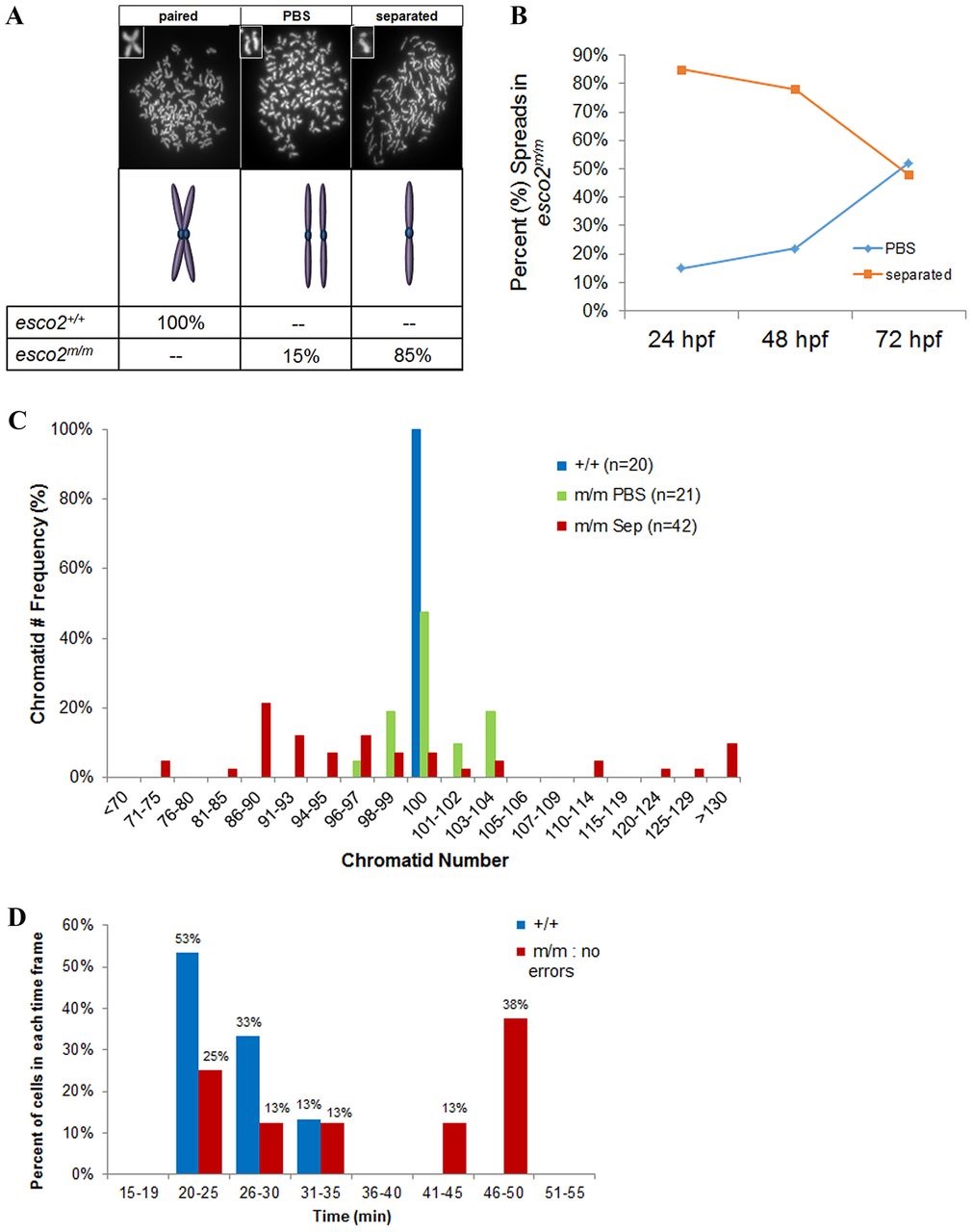
Fig. 6
Most cells of the esco2m/m embryo display complete cohesion loss, although some cells display mild cohesion defects, mild aneuploidy and almost normal mitotic transition. (A-C) Metaphase chromosome spreads from pooled (10-12 embryos) esco2+/+ (AB) and esco2m/m embryos display three key categories: ‘paired’, ‘paired but separated’ (PBS) and ‘separated’. (A) Percentage distribution of spread categories (n≥20 spreads/genotype) from pooled esco2+/+ and esco2m/m 24-hpf embryos. Insets in chromosome spreads are higher-magnified versions of the observed categories. If mixed categories were observed in the same spreads, they were counted toward the category in which the most prevalent phenotype was observed. (B) Frequency of PBS and separated spread categories from pooled esco2m/m at 24, 48 and 72hpf (n≥20 spreads/time-point). (C) Frequency of chromatid number within a spread categorized to be either the ‘paired but separated’ (PBS) or ‘separated’ phenotype from 24-hpf pooled esco2m/m mutants. Chart also contains frequency of chromatid number from esco2+/+ as a control. (D) Division time from NEB to NER of cells from esco2+/+ embryos, or cells divisions deemed ‘without error’ from esco2m/m embryo time-lapse videos.

