Image
Figure Caption
Fig. S4
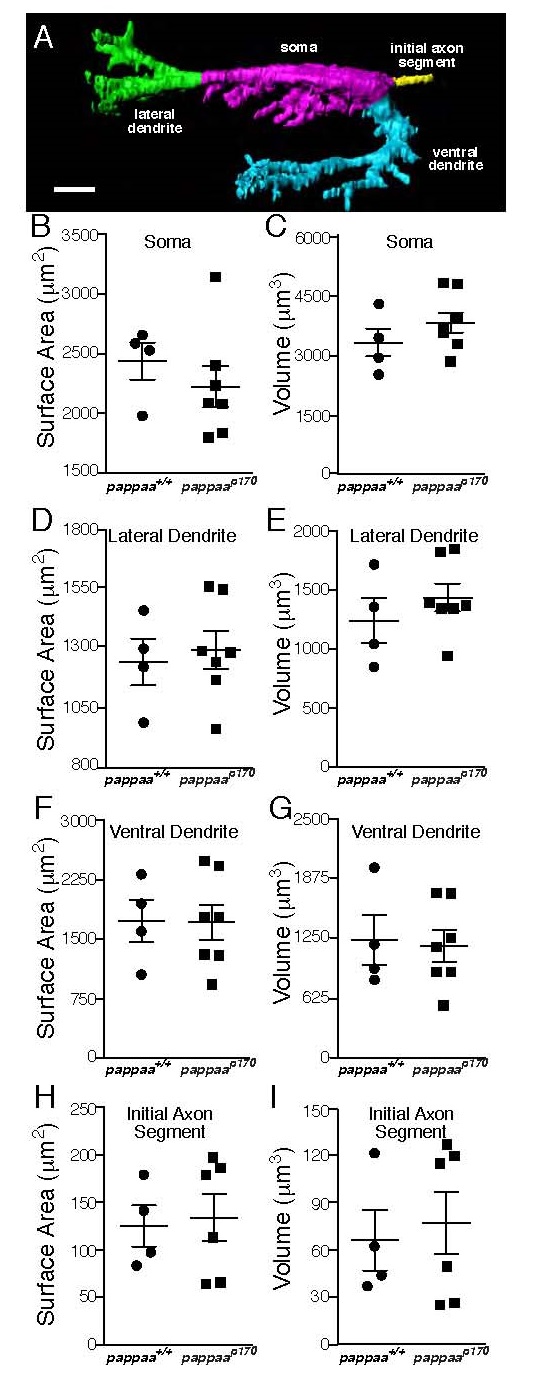
Mauthner neuron morphology (related to Figure 3).
(A) 3D reconstruction of Mauthner neuron of pappaa+/+ larva showing soma, lateral and ventral dendrites, and initial axon segment. Scale bar = 10µm. Quantification of Mauthner neuron morphology includes: mean surface area of soma (B), lateral dendrite (D), ventral dendrite (F), and initial axon segment (H) and mean volume of soma (C), lateral dendrite (E), ventral dendrite (G), and initial axon segment (I). Each mark indicates a Mauthner neuron from a single larva. Error bars denote SEM. No significant differences between pappaa+/+ and pappaap170 larvae.
Figure Data
Acknowledgments
This image is the copyrighted work of the attributed author or publisher, and
ZFIN has permission only to display this image to its users.
Additional permissions should be obtained from the applicable author or publisher of the image.
Full text @ Neuron

