Fig. 2
pappaa Expression in Neurons of the Acoustic Startle Circuit
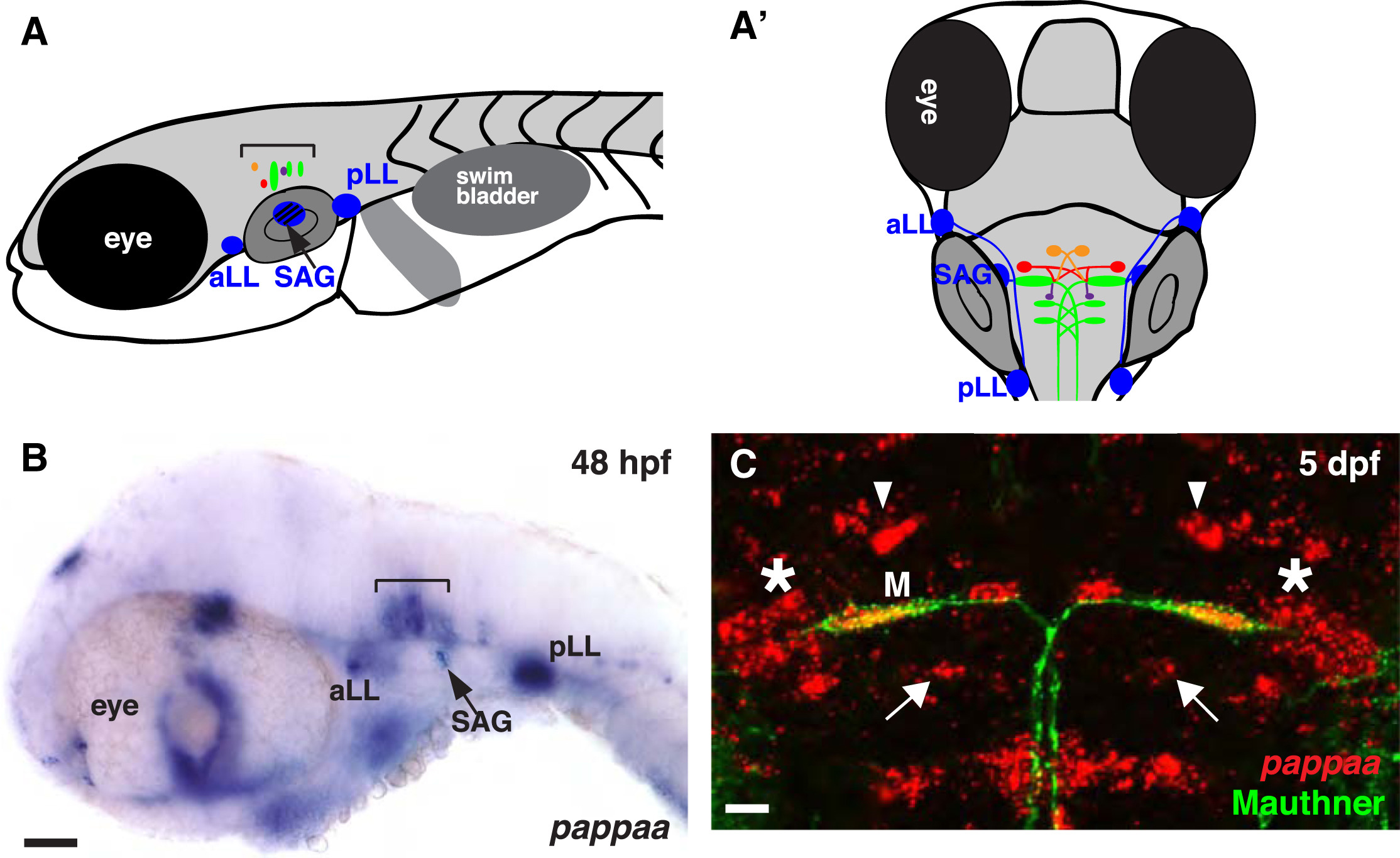
(A and A′) Schematic representation of acoustic startle circuit at larval stage from a lateral (A) and dorsal (A′) perspective. The acoustic startle circuit includes cranial ganglion (blue), Mauthner neuron and homologs (green), spiral fiber neurons (orange), passive hyperpolarizing (PHP) neurons (red), and feedback inhibitory neurons (purple).
(B and C) In situ hybridization for pappaa at 48 hpf (B, purple) and 5 dpf (C, red). Brackets (A and B) mark site of hindbrain neurons controlling startle behavior. (C) Dorsal view, anterior to the top. pappaa mRNA in red, Mauthner neuron (M) in green. Arrowheads mark site of spiral fiber neurons, asterisks mark position of PHP neurons, and arrows indicate location of feedback inhibitory neurons. SAG, statoacoustic ganglion; aLL, anterior lateral line ganglion; pLL, posterior lateral line ganglion; M, Mauthner. Scale bars = 50 µm (B) and 10 µm (C).

