
|
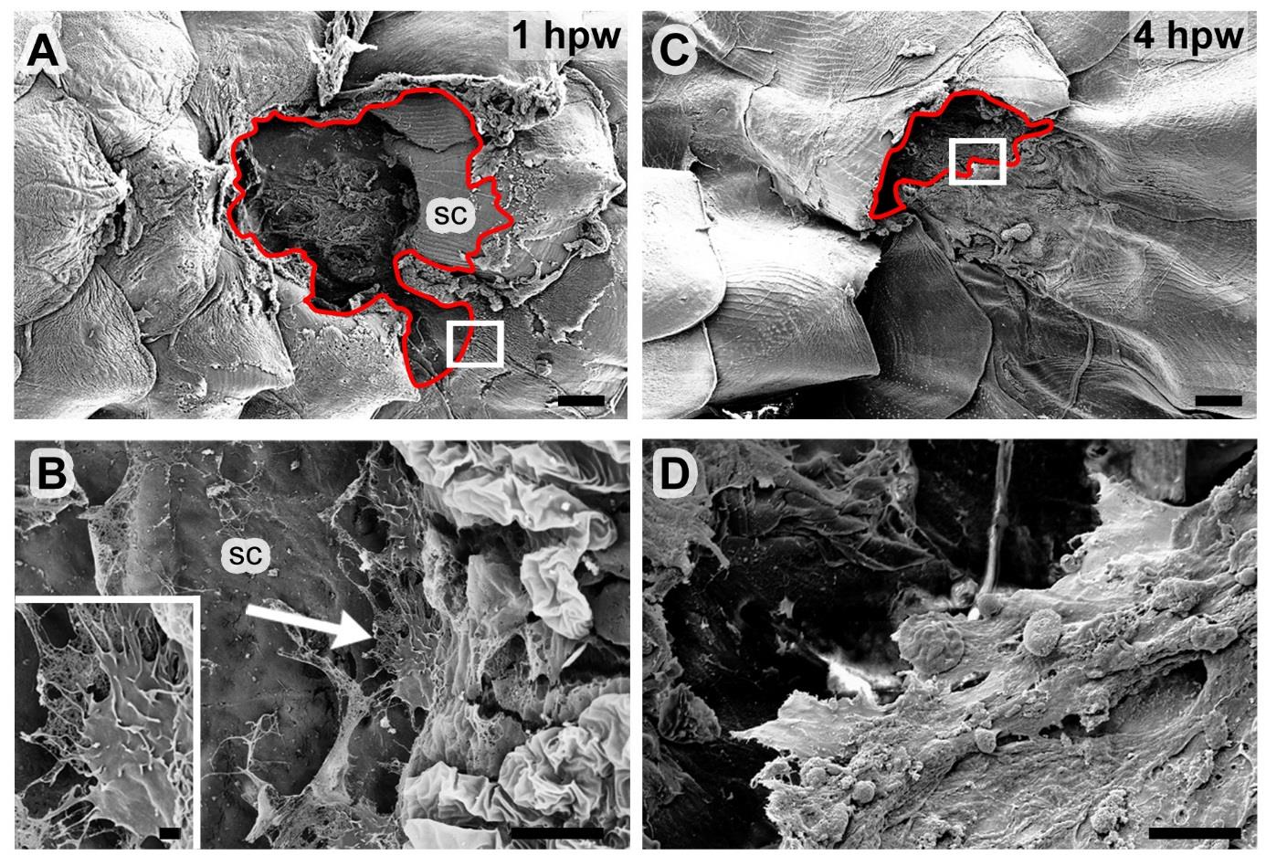
Fig. S2
LE keratinocytes of full-thickness wounds only display protrusive activity when positioned on naked scale remnants. All panels show SEM micrographs of 1 mm full-thickness wounds; (B,D) show magnified views of regions boxed in (A,C), inset in (B) shows magnified view of cellular protrusion indicated by arrow in (B). In (A,C), wound edges / leading edges of epidermal tongues are marked by red lines. Leading edge cells only extend protrusions in regions where the substrate allows, for example, on remnants of a damaged scale (A,B). In contrast, re-epithelializing epidermis covering the denuded muscle layer at the centre of the wound does not seem to be directly attached to a migration substrate, and the LE cells do not show distinct protrusions (C,D). Abbreviation: sc, scale. Scale bars: A,C = 200 µm; B,D = 10 µm; inset in B = 1 µm.