Fig. 6
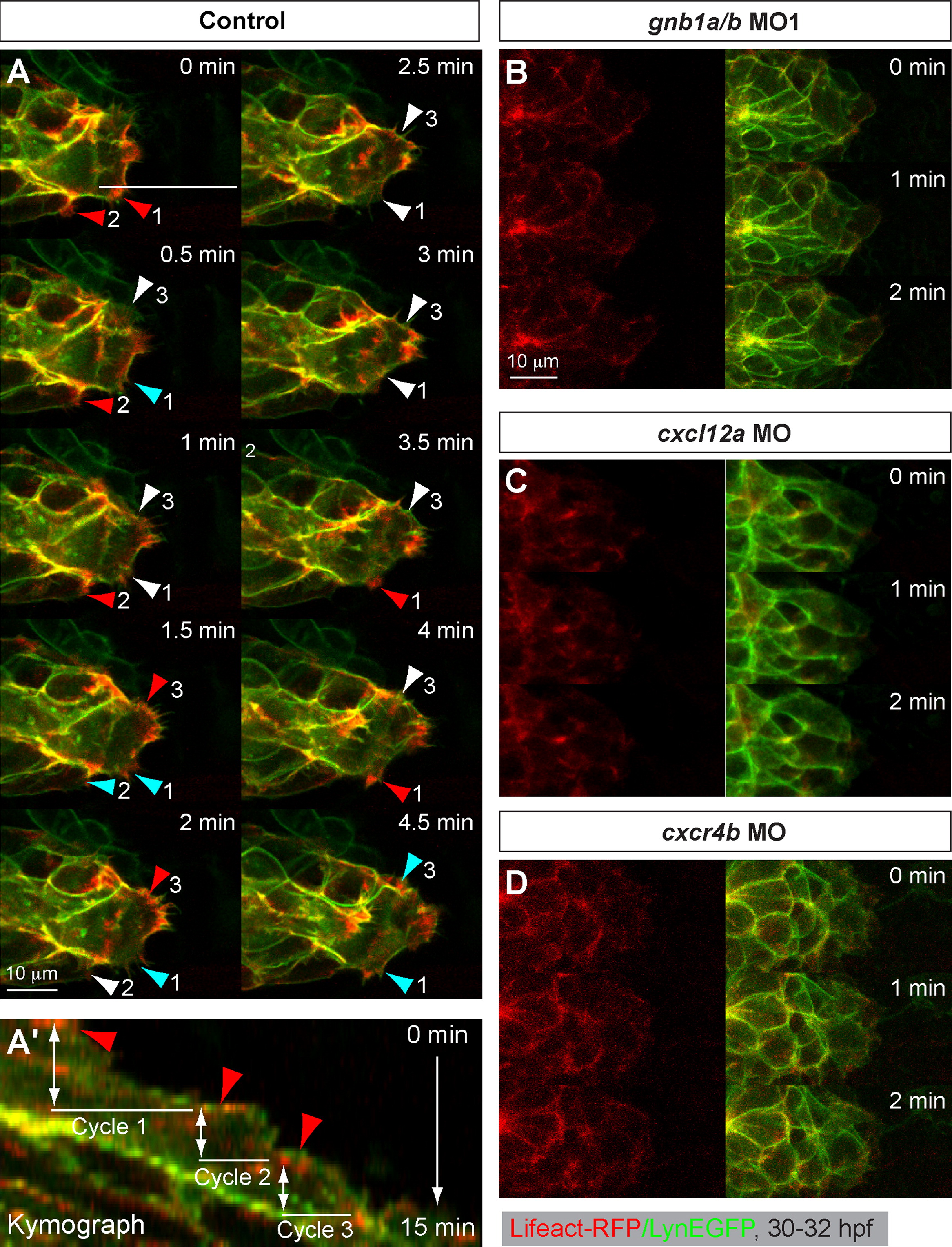
Gβ1 signaling regulates actin dynamics in the leader cells of the pLLP. Confocal time-lapse movies taken on Tg(-135bpcxcr4b:lifeact-RFP)/Tg(-8.0cldnb:lynEGFP) double-transgenic embryos at 30–32 hpf, in which actin cytoskeleton dynamics were revealed by Lifeact-RFP labeling, and pLLP cell membrane by membrane-bound EGFP. (A-A′) (A) Montage images of the control pLLP leader cells from a 4.5-min confocal time-lapse movie. A few protrusion areas (arrowheads) were highlighted (red: high actin labeling, Cyan: decreased actin labeling, and white: faint or no actin labeling). Numbers follow the same area. A 19.5-min time-lapse recording is shown in Movie 9. (A′) The kymograph image was generated from 15-min movie along the line shown in the snapshot at 0 min time point, showing the relative positions of Lifeact-RFP and LynEGFP labeling. A few cycles of association and dissociation of Lifeact-RFP enrichment (arrowheads) with GFP were shown. (B, D) Snapshots of the leading region of the pLLP of gnb1a/b MO- (B, Movies 10), cxcl12a MO- (C) or cxcr4b MO- (D) injected embryos.
Reprinted from Developmental Biology, 385(2), Xu, H., Ye, D., Behra, M., Burgess, S., Chen, S., and Lin, F., Gbeta1 controls collective cell migration by regulating the protrusive activity of leader cells in the posterior lateral line primordium, 316-27, Copyright (2014) with permission from Elsevier. Full text @ Dev. Biol.

