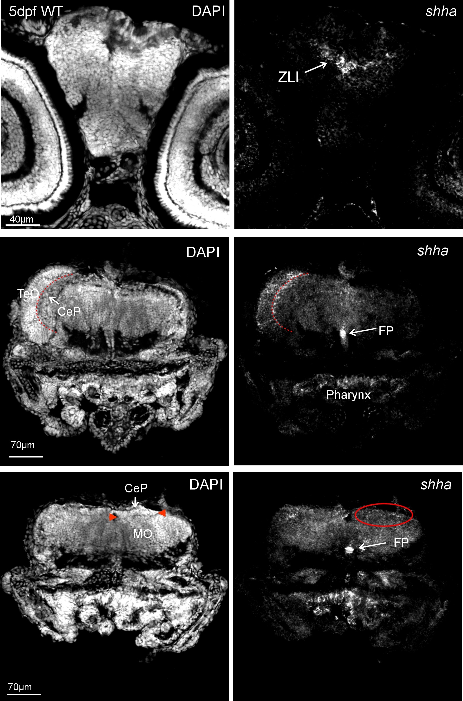
Fig. S2
Zebrafish (wild type, WT) brain section in-situ hybridized for shha at 5 dpf and analyed with confocal microscopy shows expected expression domains (right panels). Left panels: corresponding DAPI pictures for anatomical identification. Upper row: level of zona limitans intrathalamica (ZLI). Middle row: level of optic tectum (TeO) and cerebellar plate (CeP; boundary shown by red stippled line) shows shha expressing cells in floor plate (FP) and cerebellar plate, as well as in pharynx. Bottom row: level of posterior cerebellar plate (boundary towards medulla oblongata, MO, indicated by red arrowheads in left panel), shha expressing cells in FP and cerebellar plate (encircled in red), as well as in pharynx.

