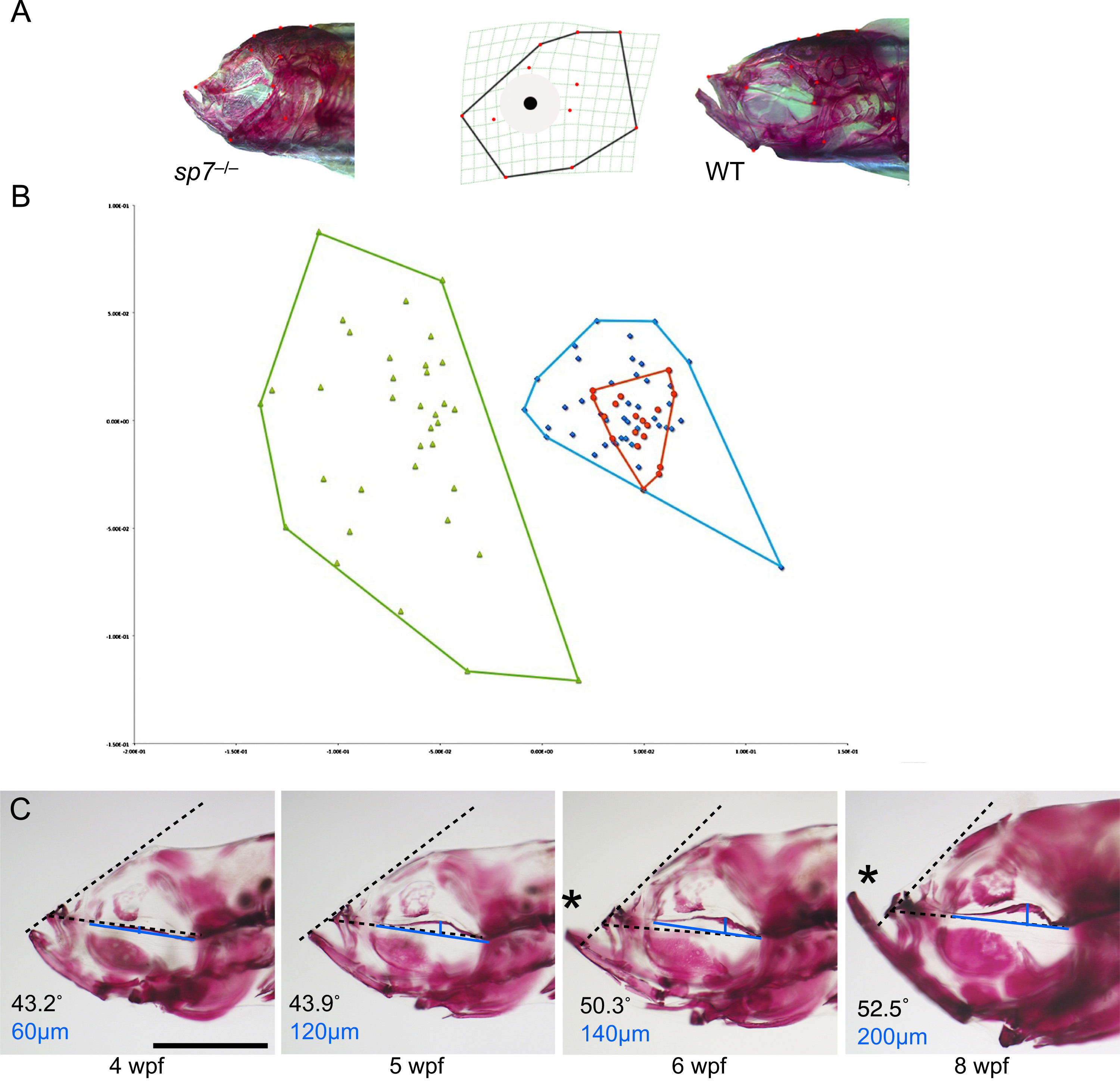
Fig. 3
Midface hypoplasia correlates with deformation of the parasphenoid. (A) 12 landmarks from the lateral view of the craniofacial skeleton of 6 wpf fish were used for morphometric analyses; representative sp7 mutant and WT fish are shown. (B) Principal component analysis separated sp7 homozygotes into a unique group (green). Heterozygotes group (blue) overlap with WT fish (red), although they are more variable (C) Alizarin red stained mutants representing progressive stages of craniofacial. development are shown in lateral view. Increasingly severe deformation of the parasphenoid is accompanied by midface hypoplasia. We quantified midface hypoplasia as the angle between the parasphenoid (dashed line drawn from the base of the parasphenoid to the anterior of the maxilla) and frontal bones (dashed line from the maxilla to frontal bones). In WT the angle remains nearly constant during development (data not shown), while in sp7 mutants the angle increases (indicated at the bottom left of each picture). Asterisks indicate protrusion of the mandible as a consequence of midface hypoplasia. To quantify the deformation of the parasphenoid, we drew a line from the caudal end of the parasphenoid to its anterior (blue line) and measured the greatest distance to the bone from this line (perpendicular blue line). Note that the distance increases with the severity of hypoplasia observed (indicated at the bottom left in blue). Scale bar is 1 mm.
Reprinted from Developmental Biology, 413(2), Kague, E., Roy, P., Asselin, G., Hu, G., Stanley, A., Albertson, C., Simonet, J., Fisher, S., Osterix/sp7 limits cranial bone initiation sites and is required for formation of sutures, 160-72, Copyright (2016) with permission from Elsevier. Full text @ Dev. Biol.

