Fig. S7
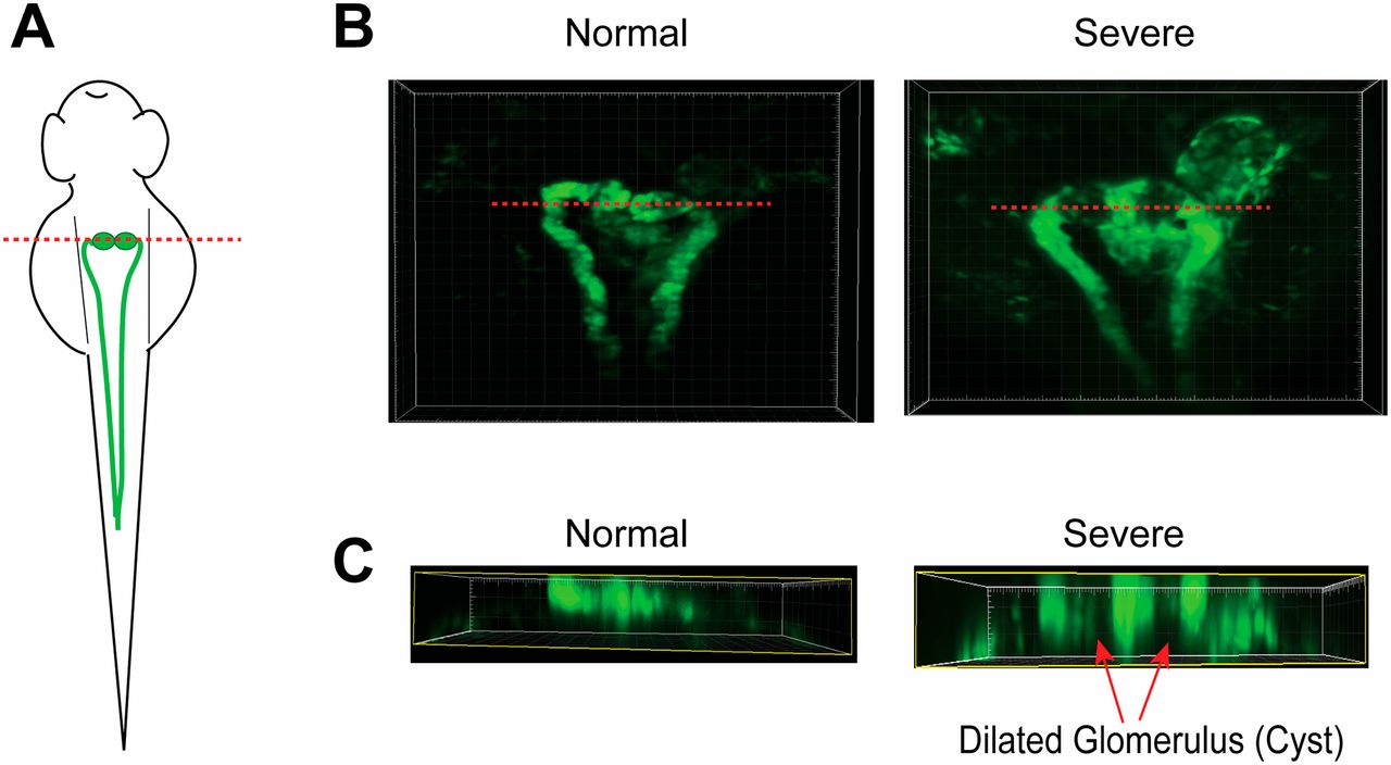
Zebrafish with severe curly tail had a diluted glomerulus (cyst) in the pronephric kidney. (A) Diagram showing zebrafish pronephric kidney at around 48 hpf. The red line indicates the position of the virtual section shown in C. (B) Confocal images of the Tg(wt1b:eGFP)li1 fish embryos with normal and severe curly tail phenotypes. GFP-positive tissues show the glomerulus and pronephric tubule. Dilation (cyst) was clearly observed in the glomerulus of the fish with the severe curly tail phenotype. (C) Virtual sections were made after 3D reconstructions of the GFP expression in the glomerulus shown in B. Red arrows indicate the dilated glomerulus (cyst) in fish with the severe curly tail phenotype.

