
|
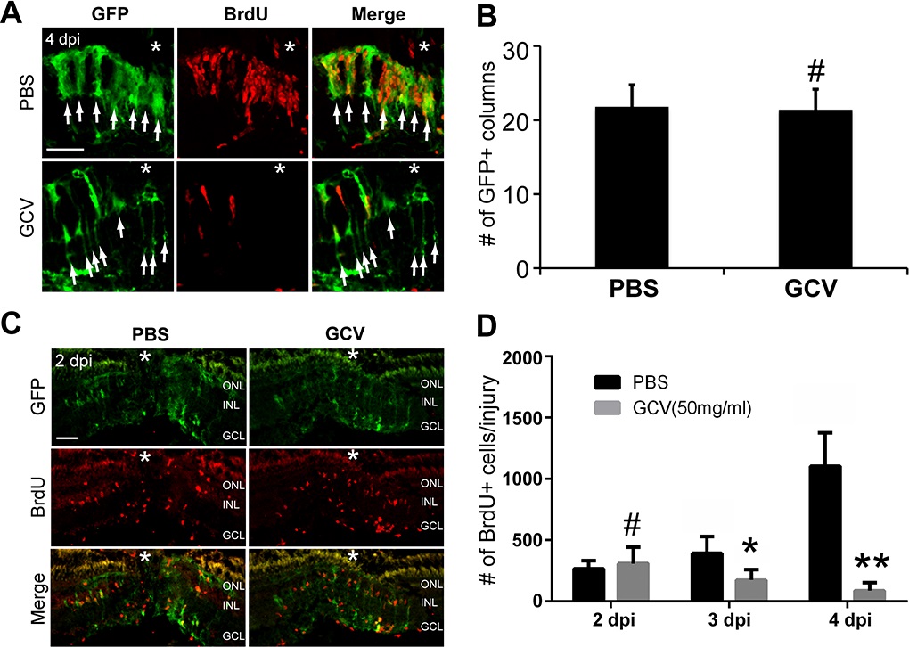
Fig. 2
Ganciclovir had no effect on Müller glia dedifferentiation and the initial MGPC formation. (A) Confocal microscopy of retinal sections after GFP and BrdU immunofluorescence shows the presence of GFP+ Müller glia near injury site in PBS- or GCV-treated retina at 4 dpi. Tg(1016tuba1a:GFP) fish were given a pulse of BrdU 3 hours before they were killed at 4 dpi. Note that there are many fewer BrdU+ MGPCs in GCV-treated retina than in control. Arrows indicate GFP+ columns. Each GFP+ column represents an activated Müller glia and its daughter progenitor cells. (B) Quantification of the number of GFP+ columns at the injury site at 4 dpi. #No significant difference, P > 0.05, n = 4. (C) Immunofluorescence shows the initial formation of MGPCs in the INL in PBS- or GCV-treated retina at 2 dpi. Tg(1016tuba1a:GFP) fish received a pulse of BrdU 3 hours before they were killed at 2 dpi. (D) Quantification of the number of BrdU+ MGPCs per injury at 2, 3, and 4 dpi in PBS- or GCV-treated retina. Fish received a pulse of BrdU 3 hours prior to killing at 2, 3, or 4 dpi. #No significant difference, P > 0.05; *P < 0.05; **P < 0.01. n = 4 for each group. Scale bars: 50 µm. The asterisks mark the injury site (needle poke).