Fig. S2
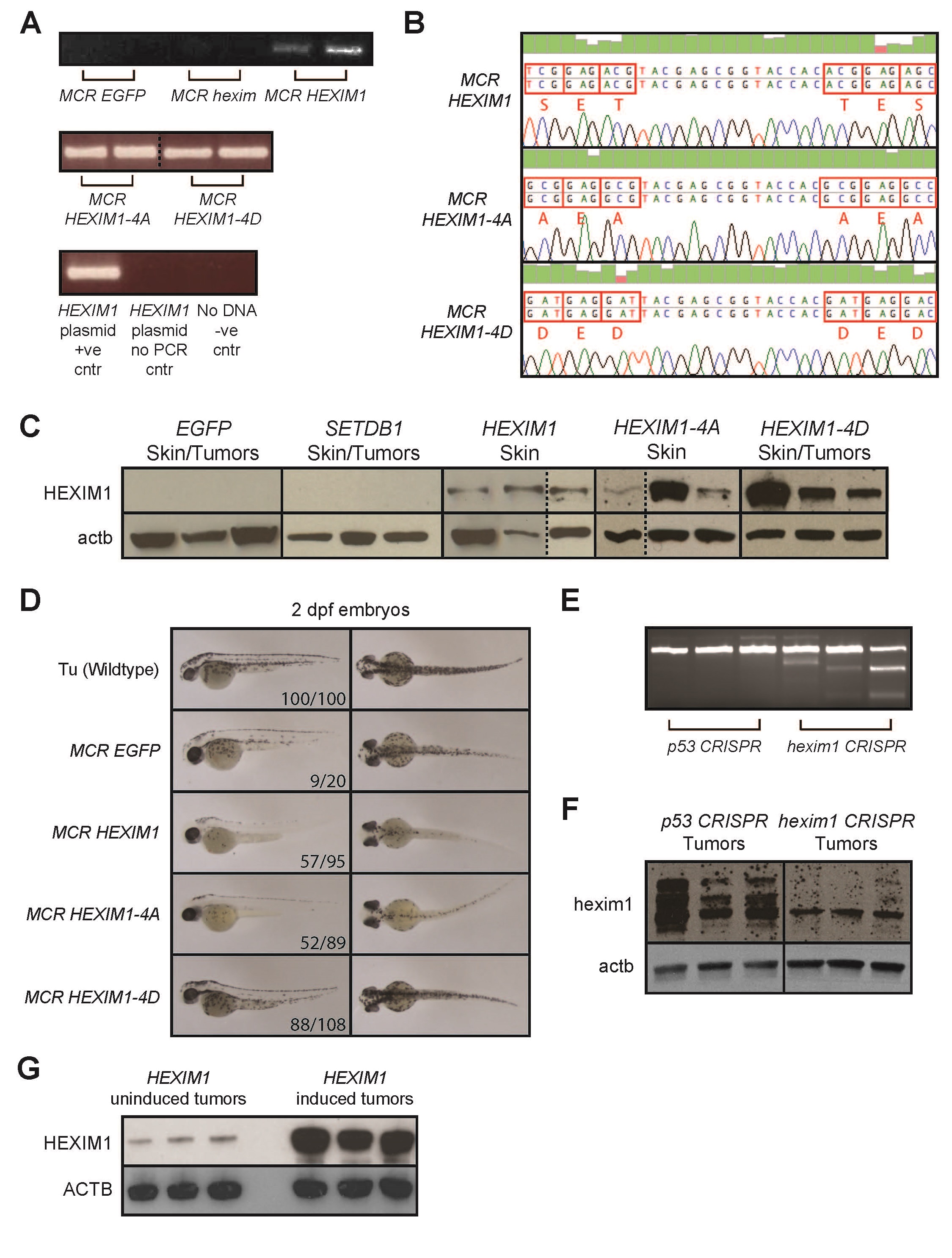
Validation of MiniCoopR HEXIM1 integration and overexpression in adult Tg(mitfa:BRAFV600E); p53-/-;mitfa-/- zebrafish, and examination of embryonic phenotypes, related to Figure 2.
(A) MiniCoopR transient and stable lines were validated by extracting genomic DNA from tail fin clips of adult zebrafish. The region of human HEXIM1 that was mutated was then amplified by PCR. PCR was run on fin clips from MCR EGFP fish, MCR hexim1 fish, a HEXIM1 plasmid, a no-DNA control and HEXIM1 plasmid with no-PCR for comparison. The dotted line denotes that samples are from different gels.
(B) PCR amplification products from (A) were sequenced to confirm strains containing WT HEXIM1, HEXIM-4A or 4D. A representative sequencing trace shows that amino acids S268, T270, T276 and S278 in human HEXIM1 were mutated to alanine in HEXIM1-4A and aspartic acid in HEXIM1-4D.
(C) Three representative adult MiniCoopR-integrated zebrafish for each strain were sacked and protein was isolated from their skin or tumors if present. Western blots for human HEXIM1 and β-actin (actb) were then performed on these samples. The dotted line denotes that samples are from different gels.
(D) EGFP, HEXIM1, HEXIM1-4A and HEXIM1-4D MiniCoopR transgenic zebrafish lines generated from the tumor onset experiment in Figure 2 were outcrossed with the Tg(mitfa:BRAFV600E);p53-/-;mitfa-/- strain (strain with no MiniCoopR integration) to give rise to offspring. Side and dorsal views of these offspring, 2 dpf zebrafish embryos, are shown. Also shown are embryos from an incross of the Tu wildtype strain for comparison.
(E) T7E1 genotyping assay for hexim1 mutation in 20 week old Tg(mitfa:BRAFV600E);p53-/-;mitfa-/- zebrafish expressing either a p53 or hexim1 gRNA under the MiniCoopR system. Cleavage bands are detected when there is a DNA heteroduplex caused by mutation.
(F) Three representative adult p53 or hexim1 CRISPR zebrafish were sacked and protein was isolated from their tumors. Western blots for hexim1 and β-actin (actb) were then performed on these samples.
(G) Protein was isolated from three representative A375-HEXIM1 mouse xenograft tumors from either the HEXIM1 uninduced (no doxycycline diet) or HEXIM1 induced (doxycycline diet) mice. Western blots for HEXIM1 and β-actin (ACTB) were performed on these samples.

