Fig. 7
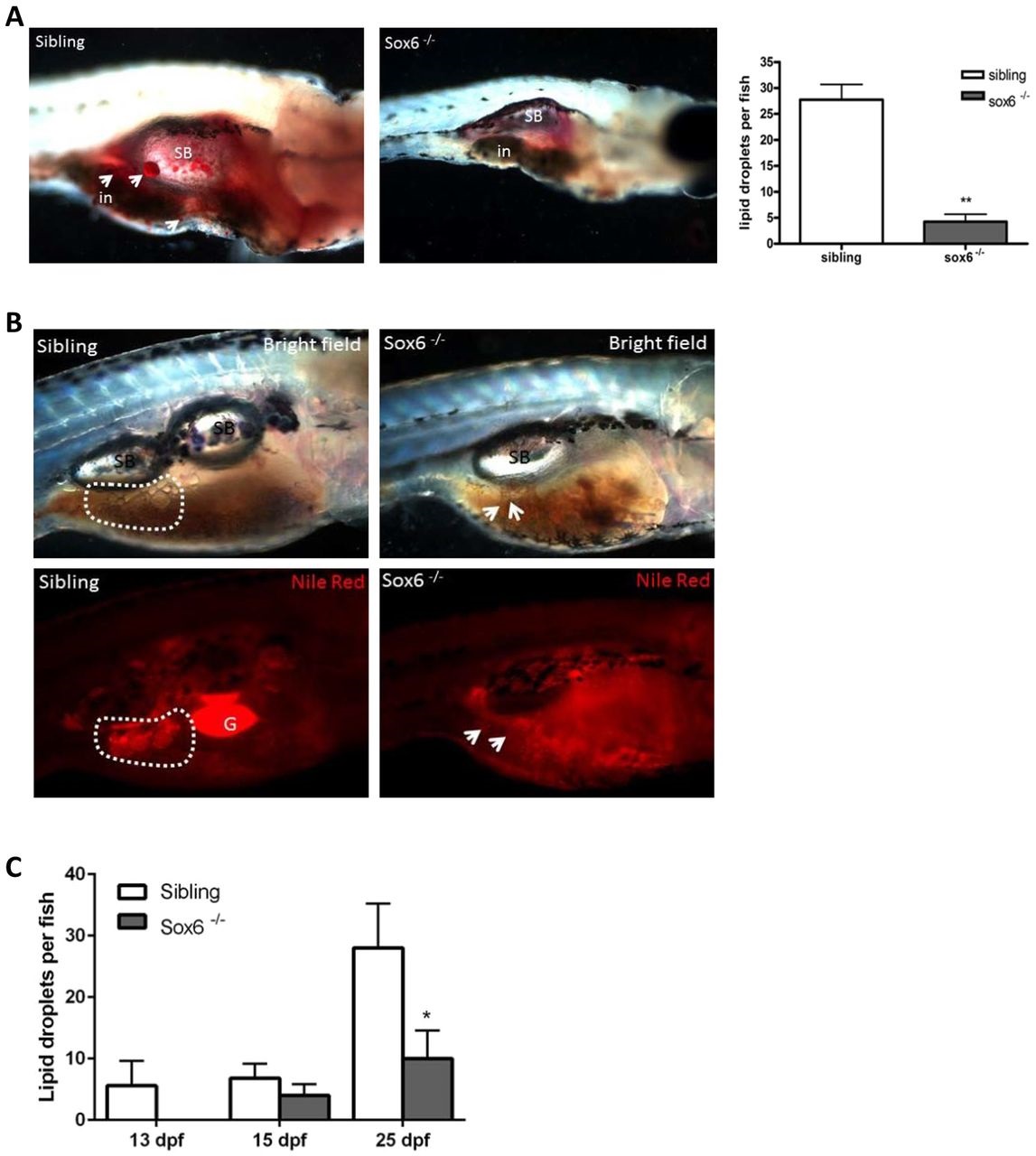
sox6 mutant zebrafish larvae exhibit decreased adipogenesis. (A) Oil Red O staining reveals localization of adipocyte neutral lipid droplets in larvae at 23days post fertilization (dpf). The arrows indicate the localization of adipocytes. The number and the size of neutral lipid droplets are decreased in sox6 mutants. Swim bladder (sb) and intestine (in) are indicated. Anterior is to the right and dorsal at the top in all images. Error bars represent s.d.; sibling n=4, mutant n=4; **P<0.01, two-tailed unpaired Student′s t-test. (B) Nile Red staining reveals that adipocyte lipid droplets are decreased in number and in size within the viscera of sox6 mutant larvae at 25dpf. Bright-field (upper panel) and corresponding fluorescence images (lower panel) are shown. Swim bladder (sb) and gall bladder (G) are indicated. The arrows indicate the localization of adipocytes (dotted line). (C) Live larvae at 13, 15 and 25dpf were stained with Nile Red. The number of lipid droplets is decreased in sox6 mutant larvae at all these developmental stages. Error bars represent s.d.; sibling n=4, mutant n=4; *P<0.05, two-tailed unpaired Student′s t-test.

