
Image
|
Figure Caption
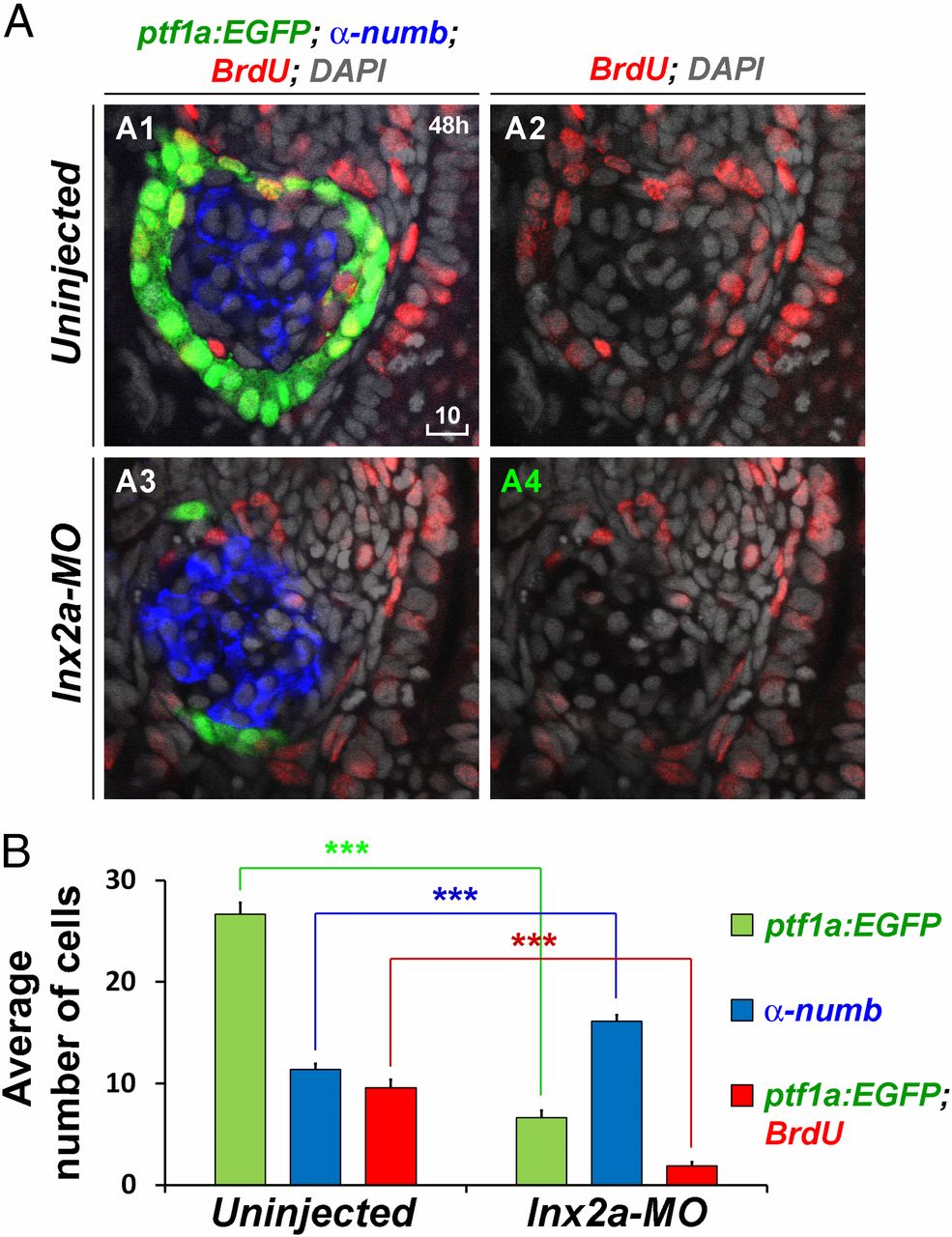
Fig. 7
lnx2a-MO injection results in impaired cell proliferation in the ventral pancreas. (A) Tg(ptf1a:EGFP) embryos were incubated in 10 mM BrdU from 46 to 47 hpf, fixed, stained with anti-BrdU and anti-Numb antibodies, and analyzed by confocal microscopy. For clarity, the same plane of the BrdU channel is shown separately in addition to the merged images. (Scale bar: 10 µm.) (B) The average number of ptf1:EGFP+, Numb+ and ptf1:EGFP+/BrdU+ double-positive cells in the pancreas of 10 uninjected and lnx2a-MO–injected embryos are shown (***P < 0.001).
Figure Data
Acknowledgments
This image is the copyrighted work of the attributed author or publisher, and
ZFIN has permission only to display this image to its users.
Additional permissions should be obtained from the applicable author or publisher of the image.
Full text @ Proc. Natl. Acad. Sci. USA