
|
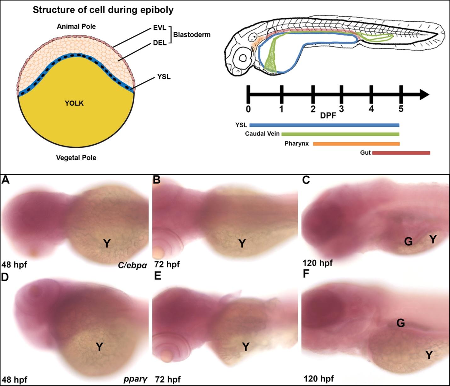
Fig. S1
The YSL separates the yolk and embryo during development and control in situ staining with the sense c/ebpα and pparγ probes (linked to Figure 1)
Left: The YSL is a multinucleated layer that forms within a few hours after fertilization that separates the yolk from the blastoderm of the embryo. Nutritional content from the yolk must pass through the YSL prior to delivery to cells. Abbreviations: EVL, enveloping layer; DEL, deep layer; YSL, yolk syncytial layer. Right: Lipid is transported from the yolk to the body at various sites during embryogenesis. Lipid must pass through the YSL (blue) to enter the body and this occurs from the early hours post fertilization through to complete yolk absorption at 5 dpf. At 24 hpf, circulation has begun and lipid can be transported by the caudal vein (green), which is in contact with the YSL, to the rest of the body. By 48 hpf, the pharynx (orange) expresses lipid metabolic genes, contains neutral lipids and is in contact with the yolk, indicating it may be involved in the lipid mobilization. At 4 dpf, lipids are present in the lumen of the gut (red), which may be processed and transported from that site. At 5 dpf, the yolk is substantially used up and feeding commences, allowing for nutritional intake to be processed in the gut. (A-C) sense c/ebpα probes showing no staining at 48, 72 and 120 hpf. (D-F) sense pparγ probes showing no staining at 48, 72 and 120 hpf. G: gut; Y: yolk sac.