
|
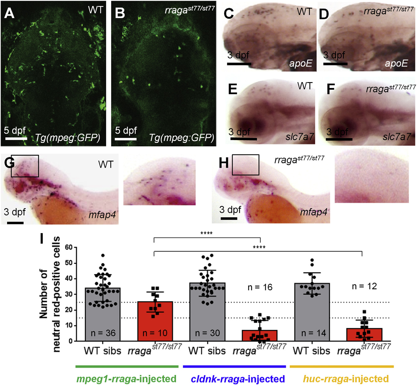
Fig. 2
rraga Acts Autonomously at an Early Stage of Microglia Development
(A and B) Reduction of microglia in rragast77/st77 mutants is detected by imaging the mpeg1:EGFP transgene in living WT (A) and mutant (B) larvae at 5 dpf. Dorsal views, anterior to the top.
(C–H) Analysis of other microglia and macrophage markers reveals reduction of microglia at 3 dpf. Probes for apoe (C and D), slc7a7 (E and F), and mfap4 (G and H) were detected by whole-mount in situ hybridization. Boxes in (G) and (H) show region magnified in the corresponding insets. Lateral views, anterior to the left.
(I) Quantification of neutral red-stained microglia in rragast77/st77 mutants after transient expression of the WT rraga coding sequence under control of regulatory sequences from mpeg1, cldnk, and huc. Only mpeg1-rraga, which drives expression in macrophages and microglia, significantly rescued microglia in the mutants. Dotted lines at 15 and 25 show weak and strong rescues, respectively (****p ≤ 0.0001).
All scale bars, 50 µm. All larvae shown were genotyped by PCR after photography (A–H) or after visually scoring neutral red phenotypes (I).