
|
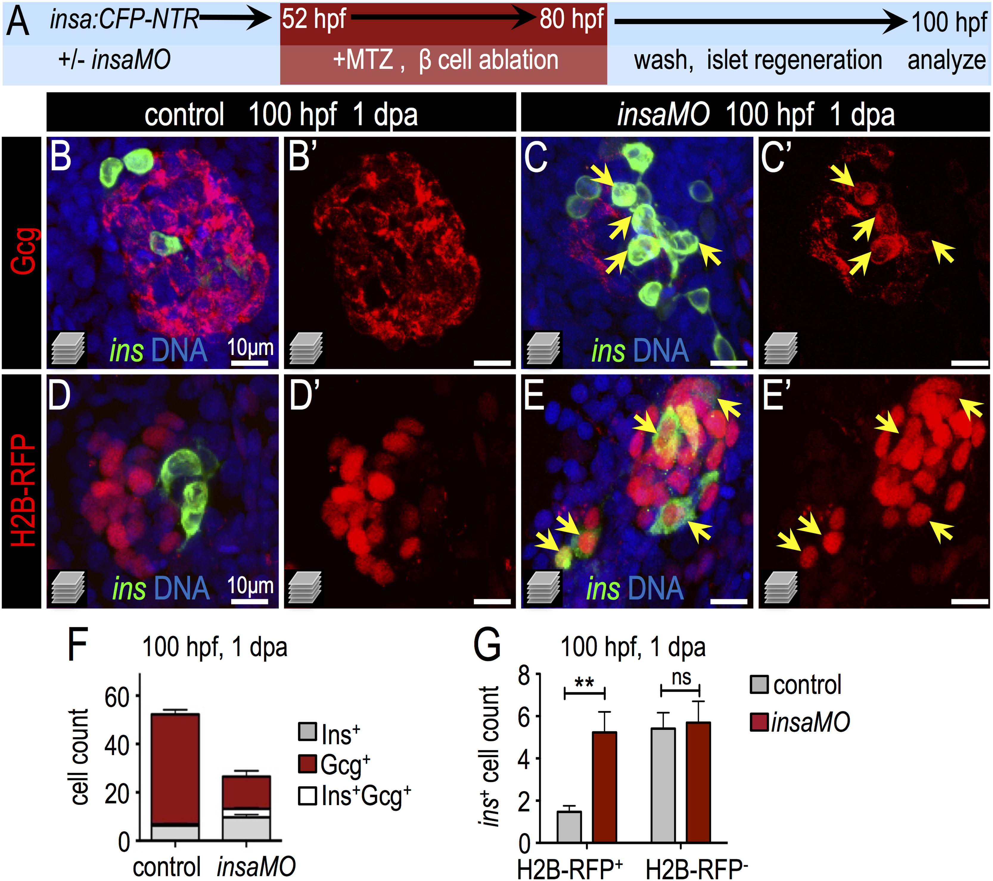
Fig. 6
Insulin knockdown enhances regeneration of β cells. (A) Schematic of β cell ablation and regeneration experiments. Control and insaMO-injected insa:CFP-NTR embryos were treated with MTZ from 52 to 80 hpf, recovered in fresh egg water for 1 day, and analyzed at ~100 hpf (1 day post ablation; dpa). (B–C′) Merged (B,C) and single channel (B′,C′) confocal projections of regenerated 100 hpf control (B,B′) and insaMO-injected (C,C′) islets immunostained for Gcg (red), CFP (β cells, green), and DNA (blue). Arrows indicate the expansion of strongly double positive Ins+ Gcg+ cells. (D–E′) Merged (D,E) and single channel (D′,E′) confocal projections of 100 hpf control (D,D′) and insaMO-injected (E,E′) islets immunostained for CFP (green) and DNA (blue). All islets expressed zygotically-injected H2B-RFP (red) to mark DBCs. Arrows indicate the expansion of double positive H2B-RFP+ Ins+ cells; this indicates conversion of non-β endocrine cells into β cells. (F) Quantification of insa+, Gcg+, and insa+ Gcg+ cells in 1 dpa control (n=17) and insaMO-injected (n=22) islets. (G) Quantification of H2B-RFP+ insa+ and H2B-RFP-insa+ regenerating β cells in 1 dpa control (n=17) and insaMO-injected (n=13) islets. Student′s t-test was used in G to determine statistical significance.
Reprinted from Developmental Biology, 409(2), Ye, L., Robertson, M.A., Mastracci, T.L., Anderson, R.M., An insulin signaling feedback loop regulates pancreas progenitor cell differentiation during islet development and regeneration, 354-69, Copyright (2016) with permission from Elsevier. Full text @ Dev. Biol.