
|
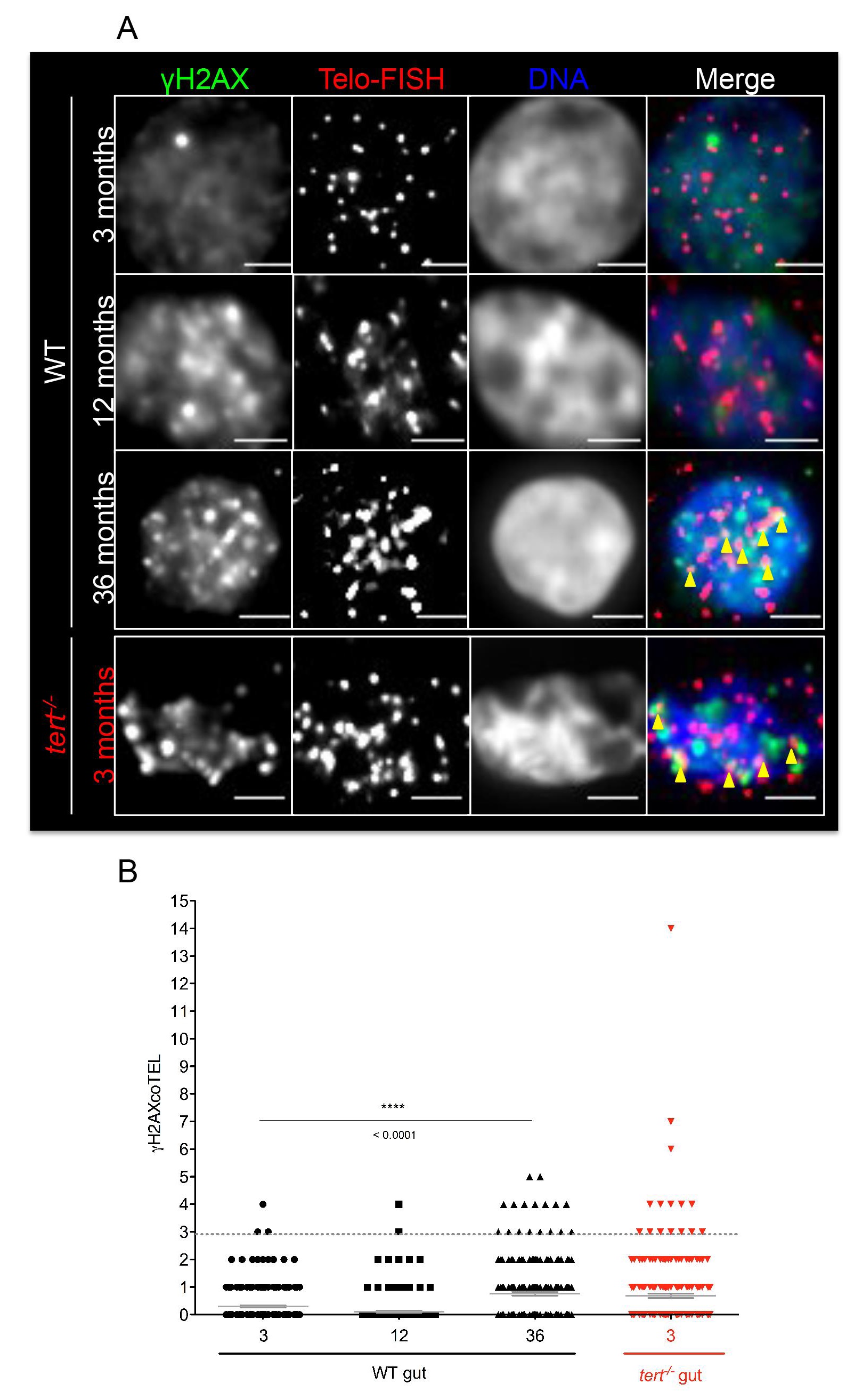
Fig. S5
The number of telomere induced foci (TIF) increases with age in the gut.A) Representative immunofluorescence staining of γ-H2AX foci (green) and telomeres by FISH (red) in cells isolated (cytospin) from the gut of 3, 12 and 36 month-old WT and 3-months tert-/-. Yellow arrowheads point to co-localization of γ-H2AX and telomere signal–TIFs. Compared to 3-months, the number of TIFs increases significantly with age in the gut of 36 months (p<0.0001) and 3 month-old tert-/- zebrafish (p<0.0001), as shown by quantifications in B). Scale bar = 1 µm. Data are represented as mean +/- SEM. N = 3 individuals, per age per time point per genotype (N = 250 cells).