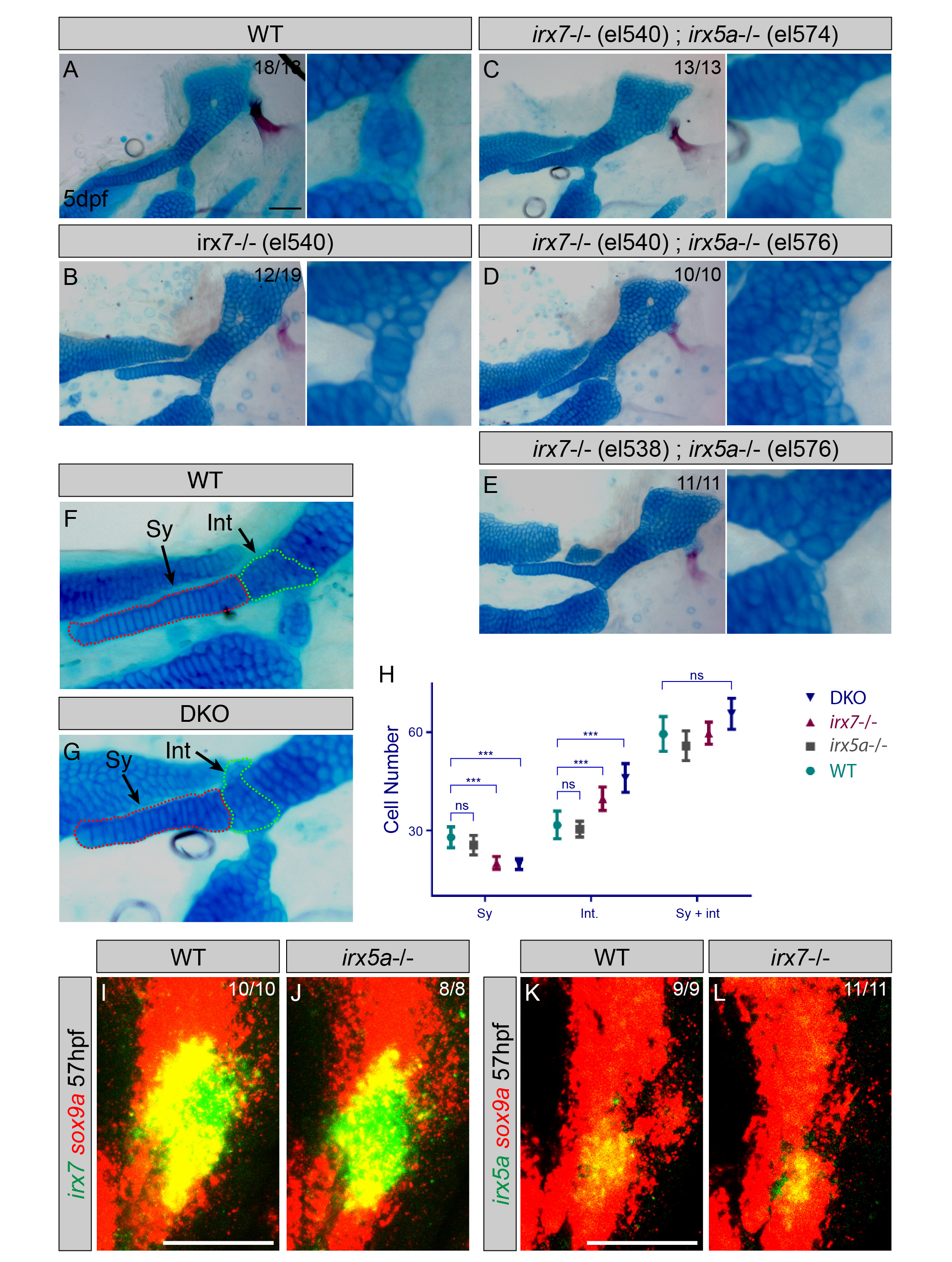
Fig. S2 Hyoid joint and symplectic cartilage defects in independent null alleles for irx7 and irx5a.
(A-E) Lateral views of hyoid skeletons (left) and magnified views of hyoid joints (right) show cartilages (Alcian Blue) and bones (Alizarin Red) at 5 dpf. Similar to the irx7el538 allele (Figure 2D), an independent irx7el540 allele shows fusions of the hyoid joint (B). All allelic combinations of irx7el538 or irx7el540 with irx5ael574 or an independent irx5ael576 allele show fully penetrant hyoid joint defects (compare C-E to Figure 2E).
(F,G) Lateral views of dissected hyoid joints and symplectic cartilage (Sy, red dashed line) in irx7el538; irx5ael574 double knock-outs (DKO) and wild-type siblings. The intermediate region between the symplectic and hyomandibular cartilages (Int, green dashed line) was outlined based on the cell shape and stacking difference from the symplectic and hyomandibula proper.
(H) Average cell counts in the Sy, Int, and combined Sy + Int regions are plotted. There were fewer cells in Sy and more cells in Int in irx7el538 and irx7el538; irx5ael574 DKO embryos compared to wild types, yet no differences in the combined numbers of cells. Cell counts were indistinguishable between wild types and irx5ael574 animals for all categories. n numbers: 8 wild type, 8 irx5a-/-, 16 irx7-/-, and 15 DKO; means are shown with 95% CI bars; *** = significant with α<0.05 using Tukey-Kramer HSD test; ns = not statistically significant.
(I-L) In situ hybridization at 57 hpf shows expression of irx7 and irx5a (green) relative to sox9a+ cartilages (red). irx7 expression is unaffected in irx5ael574 mutants, and irx5a expression is unaffected in irx7el538 mutants. Thus, at least at the level of irx7 and irx5a, there is no genetic compensation in the respective mutants. Numbers indicate proportion of animals showing the displayed phenotype. Scale bar = 50 µM.
Reprinted from Developmental Cell, 35, Askary, A., Mork, L., Paul, S., He, X., Izuhara, A.K., Gopalakrishnan, S., Ichida, J.K., McMahon, A.P., Dabizljevic, S., Dale, R., Mariani, F.V., Crump, J.G., Iroquois Proteins Promote Skeletal Joint Formation by Maintaining Chondrocytes in an Immature State, 358-65, Copyright (2015) with permission from Elsevier. Full text @ Dev. Cell

